%61%6e%74%69%63%61%6e%63%65%72
-
 KAIST Makes Cancer Cells Send Out Their Own Danger Signal — Delivering Immunotherapy and Gene Therapy in a Single Nanoparticle Platform
Cancer cells survive by hiding from the immune system's surveillance. A KAIST research team has developed a new anticancer platform that makes cancer cells send out their own danger signal—prompting immune cells to attack—while simultaneously delivering gene therapy. The approach is expected to offer a new treatment strategy that combines cancer immunotherapy and gene therapy in a single nanoparticle.
Immunogenic cell death (ICD) is a process in which dying cancer cells send danger signals to nearby immune cells, prompting them to attack. A polypeptide is a polymer made of a long chain of amino acids.
KAIST (President Choongsik Bae) announced on 28th of July that a team led by Professor Yeu-Chun Kim from the Department of Chemical and Biomolecular Engineering has developed a "helical polypeptide nanoparticle" platform that induces severe stress inside cancer cells to trigger immunogenic cell death, while also delivering a range of gene therapeutics into the cells.
The body's immune cells effectively eliminate external invaders such as viruses and bacteria, but cancer cells evade immune surveillance through a variety of immune-escape strategies. This failure of immune cells to recognize cancer cells as a threat has long been one of the biggest limitations in cancer treatment.
Recently, researchers have been actively exploring the use of nanoparticles to deliver drugs and genes to cancer cells and activate immune responses. However, it has not been clearly established which properties of nanomaterials actually induce cellular stress and activate antitumor immune responses.
By comparing and analyzing a range of nanoparticles, the team confirmed that not just the chemical composition, but the helical, coiled shape of the nanomaterial is a key factor determining therapeutic efficacy. In particular, when a positively charged quaternary amine—a chemical structure that binds readily to cell membranes—was combined with a helical structure, the particle could penetrate the cell membrane like a screw and enter cancer cells with ease. By contrast, particles with the same chemical composition but lacking the helical coil barely entered cells at all and failed to induce an immune response.
The helical nanoparticles developed by the team preferentially seek out and penetrate cancer cells, which have different membrane electrical properties from normal cells. Once inside, the particles disrupt the membranes of mitochondria—the cell's energy-producing organelles—and other organelles, subjecting the cancer cell to severe stress.
Under this extreme stress, the dying cancer cell releases damage-associated molecular patterns (DAMPs)—distress signals indicating "a dangerous cell is here"—into the surrounding environment. Immune cells that detect these signals recognize the previously hidden cancer cell as a threat and begin their attack. In effect, the cancer cell is made to broadcast its own location to the immune system.
The nanoparticle does more than trigger an immune response—it also functions as a carrier for gene therapeutics. Messenger RNA (mRNA), which carries the genetic information for protein synthesis, and small interfering RNA (siRNA), which suppresses the expression of specific genes, are both typically difficult to deliver into cells. The team's nanoparticles, however, delivered these molecules effectively into the cytoplasm.
The researchers also introduced guanidinium, a chemical functional group that binds strongly to genetic material, at an optimized ratio, enabling the particles to remain stable in the bloodstream while delivering gene therapeutics effectively.
In mouse models of melanoma and colorectal cancer, the team loaded the helical nanoparticles with siRNA targeting PD-L1 (Programmed Death-Ligand 1), an immune-evasion protein, and administered them. Tumor growth was suppressed by approximately 70–80%, and a marked increase was observed in cytotoxic T cells—which directly attack cancer cells—infiltrating the tumor, indicating a substantial boost in antitumor immune response.
"This study presents a new anticancer platform in which the nanomaterial does more than simply deliver a therapeutic agent—it drives cancer cells to trigger their own immune response," said Professor Yeu-Chun Kim. He added that the platform is expected to contribute to the development of next-generation treatments combining cancer immunotherapy and gene therapy.
Dr. Susam Lee, the paper's first author, added, "We showed that it is not just the composition of the nanomaterial but the helical structure itself that is the key factor determining therapeutic efficacy." He said he hopes the findings will serve as a new benchmark for designing next-generation immuno-oncology nanomaterials.
The study was published online in Biomaterials, a leading international journal in the field of biomaterials, on May 28, 2026.
Paper title: Helical quaternary amine polypeptide programs membrane stress to drive immunogenic cell death and cytosolic gene delivery for cancer immunotherapy
DOI: 10.1016/j.biomaterials.2026.124337
This work was supported by the National Research Foundation of Korea (NRF) grants funded by the Korean government (MSIT), the Biomedical Global Talent Nurturing Program of the Korea Health Industry Development Institute (KHIDI), funded by the Ministry of Health & Welfare, Republic of Korea (RS-2025-25459605 to Susam Lee) and the Korea Basic Science Institute (National research Facilities and Equipment Center).
2026.07.28 View 3608
-
 KAIST identifies a molecular “switch” that activates cell growth signaling, suggesting a potential basis for next-generation anticancer therapy
Cells carry their own growth switches. When enough nutrients—amino acids in particular—are available, cells flip this switch on and begin to grow. Researchers at KAIST and Yonsei University have now uncovered the molecular mechanism by which amino acid signals activate this cellular growth switch. The findings are expected to open a new avenue for anticancer therapies that target abnormal growth signaling in tumor cells.
KAIST (President Choongsik Bae) announced on July 26 that a research team led by Professors Hee-Sung Park and Jin Young Kang from the Department of Chemistry, working with Professor Sunghoon Kim's team from Yonsei University, has identified a molecular mechanism that links amino acid stimulation to mTORC1-dependent growth signaling.
Cells continually monitor whether enough amino acids—the basic building blocks of proteins—are available in their surroundings, and adjust their growth, protein synthesis, and energy use accordingly. Central to this process is mTORC1 (mammalian Target of Rapamycin Complex 1), a protein complex that functions as the cell's growth switch.
mTORC1 promotes cell growth, protein synthesis, and metabolism when nutrients and energy are abundant. But when mTORC1 becomes excessively active, cells can grow and proliferate beyond what is needed—a pattern of dysregulation observed in numerous cancers. For this reason, mTORC1 has long been considered a prime target for anticancer drug development. Exactly how cells detect external nutrient cues and translate them into mTORC1 activation, however, has remained incompletely understood.
The research team focused on the multi-tRNA synthetase complex (MSC), a large protein assembly composed of multiple aminoacyl-tRNA synthetases and scaffold proteins. While aminoacyl-tRNA synthetases are best known for their essential role in protein synthesis – attaching specific amino acids to their cognate tRNAs – the team showed that, in response to amino acid stimulation the MSC releases LARS1, thereby linking nutrient availability to growth signaling.
The key player within the MSC turned out to be a protein called LARS1 (leucyl-tRNA synthetase 1), an enzyme that attaches leucine to its corresponding tRNA and also functions as an intracellular leucine sensor. When cells receive a signal that nutrients are sufficient, LARS1 undergoes phosphorylation—a modification in which a small chemical tag is attached to a protein, altering its function or binding behavior.
The relationship can be pictured this way: the MSC is a control center where multiple proteins wait on standby, and LARS1 is the field agent dispatched to flip on the growth switch. When nutrients become abundant, LARS1 receives a phosphorylation "deployment signal," dissociates from IARS1, the protein that anchors LARS1 to the MSC, and is thereby released from the complex. The freed LARS1 then goes on to activate mTORC1.
In other words, when nutrients are scarce, LARS1 stays bound within the MSC and the growth signal remains off. Once nutrients become sufficient, LARS1 is released from the MSC and switches on mTORC1.
To investigate the structural basis of this process, the team used cryo-electron microscopy (cryo-EM), a technique that visualizes protein complexes in three dimensions in near-atomic resolution by rapidly freezing samples at extremely low temperatures. This allowed the researchers to determine how LARS1 and IARS1 bind to each other and to structurally explain how phosphorylation could disrupt their interaction.
The results showed that LARS1 and IARS1 are normally bound tightly, but amino acid stimulation induces the phosphorylation of LARS1, weakening its interaction with IARS1. This allows LARS1 to dissociate from the MSC and activate mTORC1.
The researchers also engineered phosphomimetic LARS1 variants—mutant proteins designed to imitate the phosphorylated state—and found that these variants substantially enhanced mTORC1 activity. This confirmed that the phosphorylation of LARS1 functions as the key molecular switch converting a nutrient signal into a cell growth signal.
The significance of this study lies in mapping, in concrete molecular detail, how cells sense amino acids and use that information to activate their growth switch. In particular, the study revealed that, upon receiving nutrient signals, the MSC—a complex involved in protein synthesis—releases its constituent protein LARS1, which then activates cellular growth signaling.
Some existing anticancer drugs work by directly inhibiting mTORC1, the cell's growth switch. However, because mTORC1 is also required for normal cellular growth and metabolism, its direct inhibition may also affect normal cells.
The research team expects that further identifying the kinase responsible for phosphorylating LARS1, along with its regulatory mechanism, could enable a more precise anticancer strategy—one that intercepts the growth signal further upstream, before it reaches mTORC1, rather than blocking mTORC1 itself.
The study was co-first-authored by Youjin Kim and Joo-Chan Kim from KAIST's Department of Chemistry and was published online in Nature Communications on June 11.
Paper title: Cryo-EM structure of the LARS1:IARS1 complex reveals a nutrient-responsive switch controlling mTORC1 signaling
DOI: https://doi.org/10.1038/s41467-026-74085-x
This work was supported by the National Research Foundation of Korea (grant nos. RS-2026-25482352 to H.S.P., RS-2024-00344154 to J.Y.K., and NRF-2021R1A3B1076605 to S.K.) and PNCC (grant no. 160183).
2026.07.27 View 4380
-
 Complex responsible for protein breakdown in cells identified using Bio TEM
Professor Ho-Min Kim
- High resolution 3D structure analysis success using Bio Transmission Electron Microscopy (TEM), a giant step towards new anticancer treatment development
- Published in Nature on May 5th
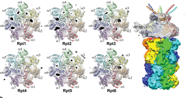
Using TEM to observe protein molecules and analysing its high resolution 3D structure is now possible. KAIST Biomedical Science and Engineering Department’s Professor Ho-Min Kim has identified the high resolution structure of proteasome complexes, which is responsible for protein breakdown in cells, using Bio TEM.
This research has been published on the world"s most prestigious journal, Nature, online on May 5th. Our body controls many cellular processes through production and degradation of proteins to maintain homeostasis. A proteasome complex acts as a garbage disposal system and degrades cellular proteins when needed for regulation, which is one of the central roles of the body.
However, a mutation in proteasome complex leads to diseases such as cancer, degenerative brain diseases, and autoimmune diseases.
Currently, the anticancer drug Velcade is used to decrease proteasome function to treat Multiple Myeloma, a form of blood cancer. Research concerning proteasome complexes for more effective anticancer drugs and treatments with fewer side effects has been taking place for more than 20 years. There have been many difficulties in understanding proteasome function through 3D structure analysis since a proteasome complex, consisting of around 30 different proteins, has a great size and complexity.
The research team used Bio TEM instead of conventionally used protein crystallography technique. The protein sample was inserted into Bio TEM, hundreds of photographs were taken from various angles, and then a high–performance computer was used to analyse its structure. Bio TEM requires a smaller sample and can analyse the complexes of great size of proteins.
Professor Ho-Min Kim said, “Identifying proteasome complex assembly process and 3D structure will increase our understanding of cellular protein degradation process and hence assist in new drug development using this knowledge.” He added, “High resolution protein structure analysis using Bio TEM, used for the first time in Korea, will enable us to observe structure analysis of large protein complexes that were difficult to approach using protein crystallography.” Professor Kim continued, “If protein crystallography technology and Bio TEM could be used together to complement one another, it would bring a great synergetic effect to protein complex 3D structure analysis research in the future.”
Professor Ho-Min Kim has conducted this research since his post-doctorate at the University of California, San Francisco, under the advice of Professor Yifan Cheng; in co-operation with Harvard University and Colorado University.
Figure 1: A picture taken by Bio TEM of open state protein sample (proteasome complex)
Figure 2: Bio TEM image analysis showing protein 3D structure
2013.05.25 View 15848