%67%65%6e%65%74%69%63
-
 Simultaneous Decoding of Genetic Maps Inside Cells... A Game Changer for Understanding Complex Human Diseases
< (Clockwise from top left) Professor Inkyung Jung (KAIST), Dr. Dongchan Yang (KAIST), Dr. Kyukwang Kim (KAIST), Dr. Yueyuan Xu (Duke University), Dr. Xiaolin Wei (Duke University), Professor Yarui Diao (Duke University) >
The origin of many diseases begins at the cellular level and involves multiple molecular interactions. However, previous methods have struggled to accurately observe changes in individual cells. Analyzing average values across thousands of cells made it challenging to detect the early signals of disease.
Our university's research team has pioneered groundbreaking technology that decodes the genetic blueprint within a cell in 3D, akin to zooming in on Earth using Google Earth. This innovation is poised to transform research into complex diseases such as cancer, dementia, and Parkinson's disease.
KAIST announced on March 4th that Professor Inkyung Jung's research team from the Department of Biological Sciences, in collaboration with Professor Yarui Diao's team at Duke University, has developed scHiCAR (single-cell Hi-C with assay for transposase-accessible chromatin and RNA sequencing). This is the world’s first ultra-high throughput & precise molecular map decoding technology that simultaneously analyzes gene expression (transcriptome), the epigenome, and the 3D genome structure within a single cell.
The key to determining a cell's state lies in how its genes operate. Genes are not simply switches that turn on and off. The destiny of a cell is determined by which genes are actually active (transcriptome), why they are active (epigenome), and within what spatial structure they operate (3D genome structure). Existing technologies required obtaining this information from different cells separately and then matching them afterward, which could lead to the distortion or omission of subtle changes.
The research team introduced ‘Trimodal Multi-omics’ technology, an integrated precision analysis method that concurrently examines these three types of genetic information within a single cell. By incorporating Artificial Intelligence (AI) analysis, they significantly enhanced accuracy and reproducibility, culminating in a unified platform that reads internal cellular genetic information akin to a ‘single 3D map.’
<Ultra-precision Single-cell Molecular Map>
Notably, the team succeeded in lowering the analysis cost to approximately $0.04 (approx. 50 KRW) per cell. Using this, they constructed a high-resolution molecular map of 1.6 million cells in mouse brain tissue. This means it is now possible to precisely identify when, where, and within what structure disease genes are turned on or off at the cellular level.
The research team applied this technology to brain tissue and the muscle regeneration process, revealing distinct gene operation principles across 22 major cell types. Notably, they successfully tracked in real-time how the 3D structure of genes dynamically changes to influence cell fate during muscle stem cell regeneration. This advancement is expected to lay a crucial foundation for developing treatment strategies for aging and incurable diseases.
<Research Result Image (AI-generated)>
Professor Inkyung Jung remarked, ‘This research transcends mere observation of cells; it opens the door to precisely reading and controlling the genomic blueprints within them. It represents a significant turning point in elucidating the developmental mechanisms of complex diseases like Parkinson's and cancer, as well as identifying target points for patient-specific new drugs.’
The study was published on February 19th in the international academic journal Nature Biotechnology (IF=46.9).
Paper Title: Trimodal single-cell profiling of transcriptome, epigenome and 3D genome in complex tissues with scHiCAR
DOI: 10.1038/s41587-026-03013-7
Meanwhile, this research was conducted with support from the Suh Kyungbae Foundation, the Samsung Science and Technology Foundation, and the Basic Research Program and Bio-Medical Technology Development Program of the National Research Foundation of Korea (Ministry of Science and ICT).
2026.03.06 View 3655
-
 A Breakthrough in Parkinson's Research: Precision Diagnosis and Treatment with AI and Optogenetics
<Research team photo (from top left) Dr. Bobae Hyeon, Professor Daesoo Kim, Director Chang-joon Lee, (right) Professor Won Do Heo>
Globally recognized figures like Muhammad Ali and Michael J. Fox have long suffered from Parkinson's disease. The disease presents a complex set of motor symptoms, including tremors, rigidity, bradykinesia, and postural instability. However, traditional diagnostic methods have struggled to sensitively detect changes in the early stages, and drugs targeting brain signal regulation have had limited clinical effectiveness.
Recently, Korean researchers successfully demonstrated the potential of a technology that integrates AI and optogenetics as a tool for precise diagnosis and therapeutic evaluation of Parkinson's disease in mice. They have also proposed a strategy for developing next-generation personalized treatments.
KAIST (President Kwang Hyung Lee) announced on the 22nd of September that a collaborative research team—comprising Professor Won Do Heo's team from the Department of Biological Sciences, Professor Daesoo Kim's team from the Department of Brain and Cognitive Sciences, and Director Chang-Jun Lee's team from the Institute for Basic Science (IBS) Center for Cognition and Sociality—achieved a preclinical research breakthrough by combining AI analysis with optogenetics. Their work simultaneously demonstrated the possibility of early and precise diagnosis and treatment in an animal model of Parkinson's disease.
The research team created a Parkinson's disease mouse model with two stages of severity. These were male mice with alpha-synuclein protein abnormalities, a standard model used to simulate human Parkinson's disease for diagnostic and therapeutic research.
In collaboration with Professor Kim's team at KAIST, they introduced AI-based 3D pose estimation for behavioral analysis. The team analyzed over 340 behavioral features—such as gait, limb movements, and tremors—from the Parkinson's mice and condensed them into a single metric: the AI-predicted Parkinson's disease score (APS).
The analysis showed that the APS exhibited a significant difference from the control group as early as two weeks after the disease was induced. It also proved more sensitive in assessing the disease's severity than traditional motor function tests. The study identified key diagnostic features, including changes in stride, asymmetrical limb movements, and chest tremors. The top 20 behavioral features included hand/foot asymmetry, changes in stride and posture, and an increase in high-frequency chest movement.
To confirm that these behavioral indicators were not just general motor decline but specific to Parkinson's, the team applied the same analysis to a mouse model of Amyotrophic Lateral Sclerosis (ALS), also known as Lou Gehrig's disease, in collaboration with Director Lee's team at IBS. Since both Parkinson's and ALS cause motor function problems, if the APS simply reflected poor motor skills, a high score should have appeared in both diseases.
However, the analysis of the ALS animal model showed that despite a decline in motor function, the mice did not exhibit the high APS seen in the Parkinson's model. Instead, their scores remained low, and their behavioral changes were distinctly different. This demonstrates that APS is directly related to specific, characteristic changes that only appear in Parkinson's disease.
For treatment, the research team used optoRET, an optogenetics technology that precisely controls neurotrophic signals with light. This technique proved effective in the animal model, leading to smoother gait and limb movements and a reduction in tremors.
Specifically, a regimen of shining light on alternate days was found to be the most effective, and it also showed a tendency to protect dopamine-producing neurons in the brain.
Professor Won Do Heo of KAIST stated, "This is the first time in the world that a preclinical framework has been implemented that connects early diagnosis, treatment evaluation, and mechanism verification of Parkinson's disease by combining AI-based behavioral analysis with optogenetics." He added, "This lays a crucial foundation for future personalized medicine and customized treatments for patients."
The study, with Dr. Bobae Hyeon, a postdoctoral researcher at the KAIST Institute for Biological Science, as the first author, was published online in the international journal Nature Communications on August 21st. Dr. Hyeon is conducting follow-up research to advance Parkinson's cell therapy at McLean Hospital, Harvard Medical School, supported by the "Global Physician-Scientist Training Program" of the Korea Health Industry Development Institute.
This research was supported by the KAIST Global Singularity Project, the Ministry of Science and ICT/National Research Foundation of Korea, the IBS Center for Cognition and Sociality, and the Ministry of Health and Welfare/Korea Health Industry Development Institute.
Paper Title: Integrating artificial intelligence and optogenetics for Parkinson's disease diagnosis and therapeutics in male mice
DOI: https://doi.org/10.1038/s41467-025-63025-w
2025.09.22 View 5658
-
 “Why are we depressed?” KAIST is identifying the cause of depression and uncovering clues for treatment
Major depressive disorder (MDD) is one of the most common psychiatric illnesses worldwide, but its molecular causes* have still not been clearly identified. A domestic research team has discovered that depression may not simply be caused by neuronal damage, but can also arise from the dysregulation of specific neural signaling pathways. In particular, they identified the molecular reason why elderly patients with depression do not respond to conventional antidepressants. This study suggests the possibility of therapeutic approaches using optogenetic technology to regulate neural signaling, and it provides clues for the development of new treatment strategies targeting the protein ‘Numb’ protein for elderly patients with depression.
*Molecular causes: explanations for the origin of a disease at the level of molecules, proteins, or genes in the brain.
KAIST (President Kwang Hyung Lee) announced on the 19th of August that a research team led by Distinguished Professor Won Do Heo of the Department of Biological Sciences at KAIST, in collaboration with forensic pathologist Minju Lee of the National Forensic Service (Director Bong Woo Lee) and Professor Seokhwi Kim of the Department of Pathology at Ajou University Medical Center (Director Sangwook Han), identified a new molecular mechanism for depression through RNA sequencing and the immunohistochemical analysis of brain tissue from patients who had committed suicide. Furthermore, they demonstrated in animal models that antidepressant effects can be restored by regulating the signaling pathway that induces neural recovery using optogenetic technology.
The research team focused on the hippocampus, the brain region responsible for memory and emotion, and in particular on the dentate gyrus (DG). The DG is the entry point of information into the hippocampus, playing a role in new memory formation, neurogenesis, and emotional regulation, and is closely linked with depression.
Using two representative mouse models for depression (the corticosterone stress model and the chronic unpredictable stress model), the team found that stress induced a striking increase in the signaling receptor FGFR1 (Fibroblast Growth Factor Receptor 1) in the DG. FGFR1 receives growth factor (FGF) signals and transmits growth and differentiation commands within cells.
Subsequently, using conditional knockout (cKO) mice in which the FGFR1 gene was deleted, the researchers revealed that the absence of FGFR1 made mice more vulnerable to stress and led them to exhibit depressive symptoms more quickly. This indicates that FGFR1 plays a critical role in proper neural regulation and stress resistance.
The team then developed an ‘optoFGFR1 system’ using optogenetics, enabling FGFR1 —essential for stress resistance—to be activated by light. They observed that activating FGFR1 in depression mouse models lacking FGFR1 restored antidepressant effects. In other words, they experimentally demonstrated that the activation of FGFR1 signaling alone could improve depressive behavior.
Surprisingly, however, in aged depression mouse models, the activation of FGFR1 signaling through the optoFGFR1 system did not yield antidepressant effects. Investigating further, the researchers found that in the aged brains, a protein called ‘Numb’ was excessively expressed and interfered with FGFR1 signaling.
Indeed, analysis of postmortem human brain tissue also showed the specific overexpression of Numb protein only in elderly patients with depression. When the researchers suppressed Numb using a gene regulatory tool (shRNA) while simultaneously activating FGFR1 signaling in mouse models, neurogenesis and behavior—previously unrecoverable—returned to normal even in aged depression models. This shows that the Numb protein acts as a “blocker” of FGFR1 signaling and is a key factor preventing the hippocampus from executing antidepressant mechanisms.
Distinguished Professor Won Do Heo of KAIST said, “This study is meaningful in that it revealed that depression may not only result from simple neuronal damage, but can also arise from the dysregulation of specific neural signaling pathways. In particular, we identified the molecular reason why antidepressants are less effective in elderly patients, and we expect this to provide a clue for the development of new therapeutic strategies targeting the Numb protein.”
He added, “Moreover, this interdisciplinary study, which combined KAIST’s expertise in neuroscience with the National Forensic Service’s forensic brain analysis technologies, is expected to serve as a bridge between basic research on psychiatric disorders and clinical applications.”
This study, led by first author Jongpil Shin, a PhD student in the Department of Biological Sciences at KAIST, was published on August 15, 2025, in the international journal Experimental & Molecular Medicine.
Paper title: “Dysregulation of FGFR1 signaling in the hippocampus facilitates depressive disorder”
DOI: https://doi.org/10.1038/s12276-025-01519-9
This research was supported by the Ministry of Science and ICT’s National Research Foundation of Korea through the ASTRA program and the Bio-Medical Technology Development project.
2025.08.19 View 6714
-
 KAIST Team Develops Optogenetic Platform for Spatiotemporal Control of Protein and mRNA Storage and Release
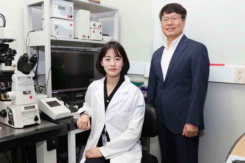
<Dr. Chaeyeon Lee, Professor Won Do Heo from Department of Biological Sciences>
A KAIST research team led by Professor Won Do Heo (Department of Biological Sciences) has developed an optogenetic platform, RELISR (REversible LIght-induced Store and Release), that enables precise spatiotemporal control over the storage and release of proteins and mRNAs in living cells and animals.
Traditional optogenetic condensate systems have been limited by their reliance on non-specific multivalent interactions, which can lead to unintended sequestration or release of endogenous molecules. RELISR overcomes these limitations by employing highly specific protein–protein (nanobody–antigen) and protein–RNA (MCP–MS2) interactions, enabling the selective and reversible compartmentalization of target proteins or mRNAs within engineered, membrane-less condensates.
In the dark, RELISR stably sequesters target molecules within condensates, physically isolating them from the cellular environment. Upon blue light stimulation, the condensates rapidly dissolve, releasing the stored proteins or mRNAs, which immediately regain their cellular functions or translational competency. This allows for reversible and rapid modulation of molecular activities in response to optical cues.
< Figure 1. Overview of the Artificial Condensate System (RELISR). The artificial condensate system, RELISR, includes "Protein-RELISR" for storing proteins and "mRNA-RELISR" for storing mRNA. These artificial condensates can be disassembled by blue light irradiation and reassembled in a dark state>
The research team demonstrated that RELISR enables temporal and spatial regulation of protein activity and mRNA translation in various cell types, including cultured neurons and mouse liver tissue. Comparative studies showed that RELISR provides more robust and reversible control of translation than previous systems based on spatial translocation.
While previous optogenetic systems such as LARIAT (Lee et al., Nature Methods, 2014) and mRNA-LARIAT (Kim et al., Nat. Cell Biol., 2019) enabled the selective sequestration of proteins or mRNAs into membrane-less condensates in response to light, they were primarily limited to the trapping phase. The RELISR platform introduced in this study establishes a new paradigm by enabling both the targeted storage of proteins and mRNAs and their rapid, light-triggered release. This approach allows researchers to not only confine molecular function on demand, but also to restore activity with precise temporal control.
< Figure 2. Cell shape change using the artificial condensate system (RELISR). A target protein, Vav2, which contributes to cell shape, was stored within the artificial condensate and then released after light irradiation. This release activated the target protein Vav2, causing a change in cell shape. It was confirmed that the storage, release, and activation of various proteins were effectively achieved>
Professor Heo stated, “RELISR is a versatile optogenetic tool that enables the precise control of protein and mRNA function at defined times and locations in living systems. We anticipate this platform will be broadly applicable for studies of cell signaling, neural circuits, and therapeutic development. Furthermore, the combination of RELISR with genome editing or tissue-targeted delivery could further expand its utility for molecular medicine.”
< Figure 3. Expression of a target mRNA using the artificial condensate system (RELISR) in mice. The genetic material for the artificial condensate system, RELISR, was injected into a living mouse. Using this system, a target mRNA was stored within the mouse's liver. Upon light irradiation, the mRNA was released, which induced the translation of a luminescent protein>
This research was conducted by first author Dr. Chaeyeon Lee, under the supervision of Professor Heo, with contributions from Dr. Daseuli Yu (co-corresponding author) and Professor YongKeun Park (co-corresponding author, Department of Physics), whose group performed quantitative imaging analyses of biophysical changes induced by RELISR in cells.
The findings were published in Nature Communications (July 7, 2025; DOI: 10.1038/s41467-025-61322-y). This work was supported by the Samsung Future Technology Foundation and the National Research Foundation of Korea.
2025.07.23 View 8193
-
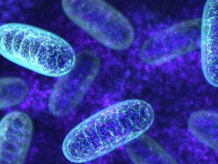 Unraveling Mitochondrial DNA Mutations in Human Cells
Throughout our lifetime, cells accumulate DNA mutations, which contribute to genetic diversity, or “mosaicism”, among cells. These genomic mutations are pivotal for the aging process and the onset of various diseases, including cancer. Mitochondria, essential cellular organelles involved in energy metabolism and apoptosis, possess their own DNA, which are susceptible to mutations. However, studies on mtDNA mutations and mosaicism have been limited due to a variety of technical challenges.
Genomic scientists from KAIST have revealed the genetic mosaicism characterized by variations in mitochondrial DNA (mtDNA) across normal human cells. This study provides fundamental insights into understanding human aging and disease onset mechanisms.
The study, “Mitochondrial DNA mosaicism in normal human somatic cells,” was published in Nature Genetics on July 22. It was led by graduate student Jisong An under the supervision of Professor Young Seok Ju from the Graduate School of Medical Science and Engineering.
Researchers from Seoul National University College of Medicine, Yonsei University College of Medicine, Korea University College of Medicine, Washington University School of Medicine National Cancer Center, Seoul National University Hospital, Gangnam Severance Hospital and KAIST faculty startup company Inocras Inc. also participated in this study.
< Figure 1. a. Flow of experiment. b. Schematic diagram illustrating the origin and dynamics of mtDNA alterations across a lifetime. >
The study involved a bioinformatic analysis of whole-genome sequences from 2,096 single cells obtained from normal human colorectal epithelial tissue, fibroblasts, and blood collected from 31 individuals. The study highlights an average of three significant mtDNA differences between cells, with approximately 6% of these variations confirmed to be inherited as heteroplasmy from the mother.
Moreover, mutations significantly increased during tumorigenesis, with some mutations contributing to instability in mitochondrial RNA. Based on these findings, the study illustrates a computational model that comprehensively elucidates the evolution of mitochondria from embryonic development to aging and tumorigenesis.
This study systematically reveals the mechanisms behind mitochondrial DNA mosaicism in normal human cells, establishing a crucial foundation for understanding the impact of mtDNA on aging and disease onset.
Professor Ju remarked, “By systematically utilizing whole-genome big data, we can illuminate previously unknown phenomena in life sciences.” He emphasized the significance of the study, adding, “For the first time, we have established a method to systematically understand mitochondrial DNA changes occurring during human embryonic development, aging, and cancer development.”
This work was supported by the National Research Foundation of Korea and the Suh Kyungbae Foundation.
2024.07.24 View 13548
-
 Overview of the 30-year history of metabolic engineering
< Distinguished Professor Sang Yup Lee from the Department of Chemical and Biomolecular Engineering at KAIST >
A research team comprised of Gi Bae Kim, Dr. So Young Choi, Dr. In Jin Cho, Da-Hee Ahn, and Distinguished Professor Sang Yup Lee from the Department of Chemical and Biomolecular Engineering at KAIST reported the 30-year history of metabolic engineering, highlighting examples of recent progress in the field and contributions to sustainability and health. Their paper “Metabolic engineering for sustainability and health” was published online in the 40th anniversary special issue of Trends in Biotechnology on January 10, 2023.
Metabolic engineering, a discipline of engineering that modifies cell phenotypes through molecular and genetic-level manipulations to improve cellular activities, has been studied since the early 1990s, and has progressed significantly over the past 30 years. In particular, metabolic engineering has enabled the engineering of microorganisms for the development of microbial cell factories capable of efficiently producing chemicals and materials as well as degrading recalcitrant contaminants.
This review article revisited how metabolic engineering has advanced over the past 30 years, from the advent of genetic engineering techniques such as recombinant DNA technologies to recent breakthroughs in systems metabolic engineering and data science aided by artificial intelligence. The research team highlighted momentous events and achievements in metabolic engineering, providing both trends and future directions in the field. Metabolic engineering’s contributions to bio-based sustainable chemicals and clean energy, health, and bioremediation were also reviewed. Finally, the research team shared their perspectives on the future challenges impacting metabolic engineering than must be overcome in order to achieve advancements in sustainability and health.
Distinguished Professor Sang Yup Lee said, “Replacing fossil resource-based chemical processes with bio-based sustainable processes for the production of chemicals, fuels, and materials using metabolic engineering has become our essential task for the future. By looking back on the 30+ years of metabolic engineering, we aimed to highlight the contributions of metabolic engineering to achieve sustainability and good health.” He added, “Metabolic engineering will play an increasingly important role as a key solution to the climate crisis, environmental pollution, food and energy shortages, and health problems in aging societies.”
< Figure: Metabolic Engineering Timeline >
2023.01.25 View 19097
-
 A Genetic Change for Achieving a Long and Healthy Life
Researchers identified a single amino acid change in the tumor suppressor protein in PTEN that extends healthy periods while maintaining longevity
Living a long, healthy life is everyone’s wish, but it is not an easy one to achieve. Many aging studies are developing strategies to increase health spans, the period of life spent with good health, without chronic diseases and disabilities. Researchers at KAIST presented new insights for improving the health span by just regulating the activity of a protein.
A research group under Professor Seung-Jae V. Lee from the Department of Biological Sciences identified a single amino acid change in the tumor suppressor protein phosphatase and tensin homolog (PTEN) that dramatically extends healthy periods while maintaining longevity. This study highlights the importance of the well-conserved tumor suppressor protein PTEN in health span regulation, which can be targeted to develop therapies for promoting healthy longevity in humans. The research was published in Nature Communications on September 24, 2021.
Insulin and insulin-like growth factor-1 (IGF-1) signaling (IIS) is one of the evolutionarily conserved aging-modulatory pathways present in life forms ranging from tiny roundworms to humans. The proper reduction of IIS leads to longevity in animals but often causes defects in multiple health parameters including impaired motility, reproduction, and growth.
The research team found that a specific amino acid change in the PTEN protein improves health status while retaining the longevity conferred by reduced IIS. They used the roundworm C. elegans, an excellent model animal that has been widely used for aging research, mainly because of its very short normal lifespan of about two to three weeks. The PTEN protein is a phosphatase that removes phosphate from lipids as well as proteins. Interestingly, the newly identified amino acid change delicately recalibrated the IIS by partially maintaining protein phosphatase activity while reducing lipid phosphatase activity.
As a result, the amino acid change in the PTEN protein maintained the activity of the longevity-promoting transcription factor Forkhead Box O (FOXO) protein while restricting the detrimental upregulation of another transcription factor, NRF2, leading to long and healthy life in animals with reduced IIS.
Professor Lee said, “Our study raises the exciting possibility of simultaneously promoting longevity and health in humans by slightly tweaking the activity of one protein, PTEN.”
This work was supported by the MInistry of Science and ICT through the National Research Foundation of Korea.
-Publication:Hae-Eun H. Park, Wooseon Hwang, Seokjin Ham, Eunah Kim, Ozlem Altintas, Sangsoon Park, Heehwa G. Son, Yujin Lee, Dongyeop Lee, Won Do Heo, and Seung-Jae V. Lee. 2021. “A PTEN variant uncouples longevity from impaired fitness in Caenorhabditis elegans with reduced insulin/IGF-1 signaling,” Nature Communications, 12(1), 5631. (https://doi.org/10.1038/s41467-021-25920-w)
-ProfileProfessor Seung-Jae V. LeeMolecular Genetics of Aging LaboratoryDepartment of Biological Sciences KAIST
2021.11.19 View 15354
-
 A Mechanism Underlying Most Common Cause of Epileptic Seizures Revealed
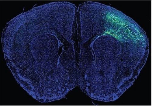
An interdisciplinary study shows that neurons carrying somatic mutations in MTOR can lead to focal epileptogenesis via non-cell-autonomous hyperexcitability of nearby nonmutated neurons
During fetal development, cells should migrate to the outer edge of the brain to form critical connections for information transfer and regulation in the body. When even a few cells fail to move to the correct location, the neurons become disorganized and this results in focal cortical dysplasia. This condition is the most common cause of seizures that cannot be controlled with medication in children and the second most common cause in adults.
Now, an interdisciplinary team studying neurogenetics, neural networks, and neurophysiology at KAIST has revealed how dysfunctions in even a small percentage of cells can cause disorder across the entire brain. They published their results on June 28 in Annals of Neurology.
The work builds on a previous finding, also by a KAIST scientists, who found that focal cortical dysplasia was caused by mutations in the cells involved in mTOR, a pathway that regulates signaling between neurons in the brain.
“Only 1 to 2% of neurons carrying mutations in the mTOR signaling pathway that regulates cell signaling in the brain have been found to include seizures in animal models of focal cortical dysplasia,” said Professor Jong-Woo Sohn from the Department of Biological Sciences. “The main challenge of this study was to explain how nearby non-mutated neurons are hyperexcitable.”
Initially, the researchers hypothesized that the mutated cells affected the number of excitatory and inhibitory synapses in all neurons, mutated or not. These neural gates can trigger or halt activity, respectively, in other neurons. Seizures are a result of extreme activity, called hyperexcitability. If the mutated cells upend the balance and result in more excitatory cells, the researchers thought, it made sense that the cells would be more susceptible to hyperexcitability and, as a result, seizures.
“Contrary to our expectations, the synaptic input balance was not changed in either the mutated or non-mutated neurons,” said Professor Jeong Ho Lee from the Graduate School of Medical Science and Engineering. “We turned our attention to a protein overproduced by mutated neurons.”
The protein is adenosine kinase, which lowers the concentration of adenosine. This naturally occurring compound is an anticonvulsant and works to relax vessels. In mice engineered to have focal cortical dysplasia, the researchers injected adenosine to replace the levels lowered by the protein. It worked and the neurons became less excitable.
“We demonstrated that augmentation of adenosine signaling could attenuate the excitability of non-mutated neurons,” said Professor Se-Bum Paik from the Department of Bio and Brain Engineering.
The effect on the non-mutated neurons was the surprising part, according to Paik. “The seizure-triggering hyperexcitability originated not in the mutation-carrying neurons, but instead in the nearby non-mutated neurons,” he said.
The mutated neurons excreted more adenosine kinase, reducing the adenosine levels in the local environment of all the cells. With less adenosine, the non-mutated neurons became hyperexcitable, leading to seizures.
“While we need further investigate into the relationship between the concentration of adenosine and the increased excitation of nearby neurons, our results support the medical use of drugs to activate adenosine signaling as a possible treatment pathway for focal cortical dysplasia,” Professor Lee said.
The Suh Kyungbae Foundation, the Korea Health Technology Research and Development Project, the Ministry of Health & Welfare, and the National Research Foundation in Korea funded this work.
-Publication:Koh, H.Y., Jang, J., Ju, S.H., Kim, R., Cho, G.-B., Kim, D.S., Sohn, J.-W., Paik, S.-B. and Lee, J.H. (2021), ‘Non–Cell Autonomous Epileptogenesis in Focal Cortical Dysplasia’ Annals of Neurology, 90: 285 299. (https://doi.org/10.1002/ana.26149)
-ProfileProfessor Jeong Ho Lee Translational Neurogenetics Labhttps://tnl.kaist.ac.kr/ Graduate School of Medical Science and Engineering KAIST
Professor Se-Bum Paik Visual System and Neural Network Laboratory http://vs.kaist.ac.kr/ Department of Bio and Brain EngineeringKAIST
Professor Jong-Woo Sohn Laboratory for Neurophysiology, https://sites.google.com/site/sohnlab2014/home Department of Biological SciencesKAIST
Dr. Hyun Yong Koh Translational Neurogenetics LabGraduate School of Medical Science and EngineeringKAIST
Dr. Jaeson Jang Ph.D.Visual System and Neural Network LaboratoryDepartment of Bio and Brain Engineering KAIST
Sang Hyeon Ju M.D.Laboratory for NeurophysiologyDepartment of Biological SciencesKAIST
2021.08.26 View 26697
-
 Rare Mutations May Have Big Impact on Schizophrenia Pathology
- Somatic mutations found only in brain cells disrupt synaptic function. -
Schizophrenia is a neurodevelopmental disorder that disrupts brain activity, producing hallucinations, delusions, and other cognitive disturbances. Researchers have long searched for genetic influences in the disease, but genetic mutations have been identified in only a small fraction—fewer than a quarter—of sequenced patients. Now a study shows that “somatic” gene mutations in brain cells could account for some of the disease’s neuropathology.
The results of the study, led by Professor Jeong Ho Lee at the Graduate School of Medical Science and Engineering in collaboration with the Stanley Medical Research Institute in the US, appeared in Biological Psychiatry.
Traditional genetic mutations, called germline mutations, occur in sperm or egg cells and are passed on to offspring by their parents. Somatic mutations, in contrast, occur in an embryo after fertilization, and they can show up throughout the body or in isolated pockets of tissues, making them much harder to detect from blood or saliva samples, which are typically used for such sequencing studies.
Recently, more-advanced genetic sequencing techniques have allowed researchers to detect somatic mutations and studies have shown that even mutations present at very low levels can have functional consequences. A previous study hinted that brain somatic mutations were associated with schizophrenia, but it was not powerful enough to cement an association between brain somatic mutations and schizophrenia.
In the current study, the researchers used deep whole-exome sequencing to determine the genetic code of all exomes, the parts of genes that encode proteins. The scientists sequenced postmortem samples from brain, liver, spleen, or heart tissue of 27 people with schizophrenia and 31 control participants allowing them to compare the sequences in the two tissues. Using a powerful analytic technique, the team identified an average of 4.9 somatic single-nucleotide variants, or mutations, in brain samples from people with schizophrenia, and 5.6 somatic single-nucleotide variants in brain samples from control subjects.
Although there were no significant quantitative differences in somatic single-nucleotide variants between schizophrenia and control tissue samples, the researchers found that the mutations in schizophrenia patients were found in genes already associated with schizophrenia. Of the germline mutations that had previously been associated with schizophrenia, the genes affected encode proteins associated with synaptic neural communication, particularly in a brain region called the dorsolateral prefrontal cortex.
In the new analysis, the researchers determined which proteins might be affected by the newly identified somatic mutations. Remarkably, a protein called GRIN2B emerged as highly affected and two patients with schizophrenia carried somatic mutations on the GRIN2B gene itself. GRIN2B is a protein component of NMDA-type glutamate receptors, which are critical for neural signaling. Faulty glutamate receptors have long been suspected of contributing to schizophrenia pathology; GRIN2B ranks among the most-studied genes in schizophrenia. The somatic mutations identified in the study had a variant allele frequency of only ~1%, indicating that the mutations were rare among brain cells as a whole. Nevertheless, they have the potential to create widespread cortical dysfunction.
Professor Lee said, “Besides the comprehensive genetic analysis of brain-only mutations in postmortem tissues from schizophrenia patients, this study experimentally showed the biological consequence of identified somatic mutations, which led to neuronal abnormalities associated with schizophrenia. Thus, this study suggests that brain somatic mutations can be a hidden major contributor to schizophrenia and provides new insights into the molecular genetic architecture of schizophrenia.
John Krystal, MD, editor of Biological Psychiatry, said of the work, "The genetics of schizophrenia has received intensive study for several decades. Now a new possibility emerges, that in some cases, mutations in the DNA of brain cells contributes to the biology of schizophrenia. Remarkably this new biology points to an old schizophrenia story: NMDA glutamate receptor dysfunction. Perhaps the path through which somatic mutations contribute to schizophrenia converges with other sources of abnormalities in glutamate signaling in this disorder."
Professor Lee and the team next want to assess the functional consequences of the somatic mutations. Because of the location of the GRIN2B mutations found in schizophrenia patients, the researchers hypothesized that they might interfere with the receptors’ localization on neurons. Experiments on the cortical neurons of mice showed that the mutations indeed disrupted the receptors’ usual localization to dendrites, the “listening” ends of neurons, which in turn prevented the formation of normal synapses in the neurons. This finding suggests that the somatic mutations could disrupt neural communication, contributing to schizophrenia pathology.
- Profile:
Professor Jeong Ho Lee
Translational Neurogenetics Laboratory ( https://tnl.kaist.ac.kr/)
The Graduate School of Medical Science and Engineering
KAIST
(END)
2021.03.11 View 13965
-
 Tinkering with Roundworm Proteins Offers Hope for Anti-aging Drugs
- The somatic nuclear protein kinase VRK-1 increases the worm’s lifespan through AMPK activation, and this mechanism can be applied to promoting human longevity, the study reveals. -
KAIST researchers have been able to dial up and down creatures’ lifespans by altering the activity of proteins found in roundworm cells that tell them to convert sugar into energy when their cellular energy is running low. Humans also have these proteins, offering up the intriguing possibilities for developing longevity-promoting drugs. These new findings were published on July 1 in Science Advances.
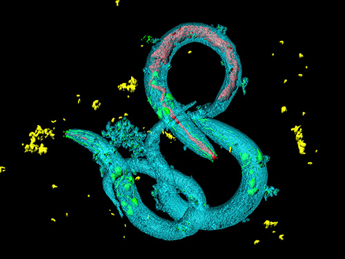
The roundworm Caenorhabditis elegans (C. elegans), a millimeter-long nematode commonly used in lab testing, enjoyed a boost in its lifespan when researchers tinkered with a couple of proteins involved in monitoring the energy use by its cells.
The proteins VRK-1 and AMPK work in tandem in roundworm cells, with the former telling the latter to get to work by sticking a phosphate molecule, composed of one phosphorus and four oxygen atoms, on it. In turn, AMPK’s role is to monitor energy levels in cells, when cellular energy is running low. In essence, VRK-1 regulates AMPK, and AMPK regulates the cellular energy status.
Using a range of different biological research tools, including introducing foreign genes into the worm, a group of researchers led by Professor Seung-Jae V. Lee from the Department of Biological Sciences at KAIST were able to dial up and down the activity of the gene that tells cells to produce the VRK-1 protein. This gene has remained pretty much unchanged throughout evolution. Most complex organisms have this same gene, including humans.
Lead author of the study Sangsoon Park and his colleagues confirmed that the overexpression, or increased production, of the VRK-1 protein boosted the lifespan of the C. elegans, which normally lives just two to three weeks, and the inhibition of VRK-1 production reduced its lifespan.
The research team found that the activity of the VRK-1-to-AMPK cellular-energy monitoring process is increased in low cellular energy status by reduced mitochondrial respiration, the set of metabolic chemical reactions that make use of the oxygen the worm breathes to convert macronutrients from food into the energy “currency” that cells spend to do everything they need to do.
It is already known that mitochondria, the energy-producing engine rooms in cells, play a crucial role in aging, and declines in the functioning of mitochondria are associated with age-related diseases. At the same time, the mild inhibition of mitochondrial respiration has been shown to promote longevity in a range of species, including flies and mammals.
When the research team performed similar tinkering with cultured human cells, they found they could also replicate this ramping up and down of the VRK-1-to-AMPK process that occurs in roundworms.
“This raises the intriguing possibility that VRK-1 also functions as a factor in governing human longevity, and so perhaps we can start developing longevity-promoting drugs that alter the activity of VRK-1,” explained Professor Lee.
At the very least, the research points us in an interesting direction for investigating new therapeutic strategies to combat metabolic disorders by targeting the modulation of VRK-1. Metabolic disorders involve the disruption of chemical reactions in the body, including diseases of the mitochondria.
But before metabolic disorder therapeutics or longevity drugs can be contemplated by scientists, further research still needs to be carried out to better understand how VRK-1 works to activate AMPK, as well as figure out the precise mechanics of how AMPK controls cellular energy.
This work was supported by the National Research Foundation (NRF), and the Ministry of Science and ICT (MSIT) of Korea.
Image credit: Seung-Jae V. LEE, KAIST.
Image usage restrictions: News organizations may use or redistribute this image, with proper attribution, as part of news coverage of this paper only.
Publication:
Park, S., et al. (2020) ‘VRK-1 extends life span by activation of AMPK via phosphorylation’. Science Advances, Volume 6. No. 27, eaaw7824. Available online at https://doi.org/10.1126/sciadv.aaw7824
Profile: Seung-Jae V. Lee, Ph.D.
Professor
seungjaevlee@kaist.ac.kr
https://sites.google.com/view/mgakaist
Molecular Genetics of Aging Laboratory
Department of Biological Sciences
Korea Advanced Institute of Science and Technology (KAIST)
https://www.kaist.ac.krDaejeon 34141, Korea
(END)
2020.07.31 View 18683
-
 Hydrogel-Based Flexible Brain-Machine Interface
The interface is easy to insert into the body when dry, but behaves ‘stealthily’ inside the brain when wet
Professor Seongjun Park’s research team and collaborators revealed a newly developed hydrogel-based flexible brain-machine interface. To study the structure of the brain or to identify and treat neurological diseases, it is crucial to develop an interface that can stimulate the brain and detect its signals in real time. However, existing neural interfaces are mechanically and chemically different from real brain tissue. This causes foreign body response and forms an insulating layer (glial scar) around the interface, which shortens its lifespan.
To solve this problem, the research team developed a ‘brain-mimicking interface’ by inserting a custom-made multifunctional fiber bundle into the hydrogel body. The device is composed not only of an optical fiber that controls specific nerve cells with light in order to perform optogenetic procedures, but it also has an electrode bundle to read brain signals and a microfluidic channel to deliver drugs to the brain.
The interface is easy to insert into the body when dry, as hydrogels become solid. But once in the body, the hydrogel will quickly absorb body fluids and resemble the properties of its surrounding tissues, thereby minimizing foreign body response.
The research team applied the device on animal models, and showed that it was possible to detect neural signals for up to six months, which is far beyond what had been previously recorded. It was also possible to conduct long-term optogenetic and behavioral experiments on freely moving mice with a significant reduction in foreign body responses such as glial and immunological activation compared to existing devices.
“This research is significant in that it was the first to utilize a hydrogel as part of a multifunctional neural interface probe, which increased its lifespan dramatically,” said Professor Park. “With our discovery, we look forward to advancements in research on neurological disorders like Alzheimer’s or Parkinson’s disease that require long-term observation.”
The research was published in Nature Communications on June 8, 2021. (Title: Adaptive and multifunctional hydrogel hybrid probes for long-term sensing and modulation of neural activity) The study was conducted jointly with an MIT research team composed of Professor Polina Anikeeva, Professor Xuanhe Zhao, and Dr. Hyunwoo Yook.
This research was supported by the National Research Foundation (NRF) grant for emerging research, Korea Medical Device Development Fund, KK-JRC Smart Project, KAIST Global Initiative Program, and Post-AI Project.
-Publication
Park, S., Yuk, H., Zhao, R. et al. Adaptive and multifunctional hydrogel hybrid probes for long-term sensing and modulation of neural activity. Nat Commun 12, 3435 (2021). https://doi.org/10.1038/s41467-021-23802-9
-Profile
Professor Seongjun Park
Bio and Neural Interfaces Laboratory
Department of Bio and Brain Engineering
KAIST
2020.07.13 View 15886
-
 Scientists Discover the Mechanism of DNA High-Order Structure Formation
(Molecular structures of Abo1 in different energy states (left), Demonstration of an Abo1-assisted histone loading onto DNA by the DNA curtain assay. )
The genetic material of our cells—DNA—exists in a high-order structure called “chromatin”. Chromatin consists of DNA wrapped around histone proteins and efficiently packs DNA into a small volume. Moreover, using a spool and thread analogy, chromatin allows DNA to be locally wound or unwound, thus enabling genes to be enclosed or exposed. The misregulation of chromatin structures results in aberrant gene expression and can ultimately lead to developmental disorders or cancers. Despite the importance of DNA high-order structures, the complexity of the underlying machinery has circumvented molecular dissection.
For the first time, molecular biologists have uncovered how one particular mechanism uses energy to ensure proper histone placement onto DNA to form chromatin. They published their results on Dec. 17 in Nature Communications.
The study focused on proteins called histone chaperones. Histone chaperones are responsible for adding and removing specific histones at specific times during the DNA packaging process. The wrong histone at the wrong time and place could result in the misregulation of gene expression or aberrant DNA replication. Thus, histone chaperones are key players in the assembly and disassembly of chromatin.
“In order to carefully control the assembly and disassembly of chromatin units, histone chaperones act as molecular escorts that prevent histone aggregation and undesired interactions,” said Professor Ji-Joon Song in the Department of Biological Sciences at KAIST. “We set out to understand how a unique histone chaperone uses chemical energy to assemble or disassemble chromatin.”
Song and his team looked to Abo1, the only known histone chaperone that utilizes cellular energy (ATP). While Abo1 is found in yeast, it has an analogous partner in other organisms, including humans, called ATAD2. Both use ATP, which is produced through a cellular process where enzymes break down a molecule’s phosphate bond. ATP energy is typically used to power other cellular processes, but it is a rare partner for histone chaperones.
“This was an interesting problem in the field because all other histone chaperones studied to date do not use ATP,” Song said.
By imaging Abo1 with a single-molecule fluorescence imaging technique known as the DNA curtain assay, the researchers could examine the protein interactions at the single-molecule level. The technique allows scientists to arrange the DNA molecules and proteins on a single layer of a microfluidic chamber and examine the layer with fluorescence microscopy.
The researchers found through real-time observation that Abo1 is ring-shaped and changes its structure to accommodate a specific histone and deposit it on DNA. Moreover, they found that the accommodating structural changes are powered by ADP.
“We discovered a mechanism by which Abo1 accommodates histone substrates, ultimately allowing it to function as a unique energy-dependent histone chaperone,” Song said. “We also found that despite looking like a protein disassembly machine, Abo1 actually loads histone substrates onto DNA to facilitate chromatin assembly.”
The researchers plan to continue exploring how energy-dependent histone chaperones bind and release histones, with the ultimate goal of developing therapeutics that can target cancer-causing misbehavior by Abo1’s analogous human counterpart, ATAD2.
-Profile
Professor Ji-Joon Song
Department of Biological Sciences KI for the BioCentury (https://kis.kaist.ac.kr/index.php?mid=KIB_O) KAIST
2020.01.07 View 19400