OT
-
 A Quick but Clingy Creepy-Crawler that will MARVEL You
Engineered by KAIST Mechanics, a quadrupedal robot climbs steel walls and crawls across metal ceilings at the fastest speed that the world has ever seen.
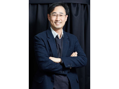
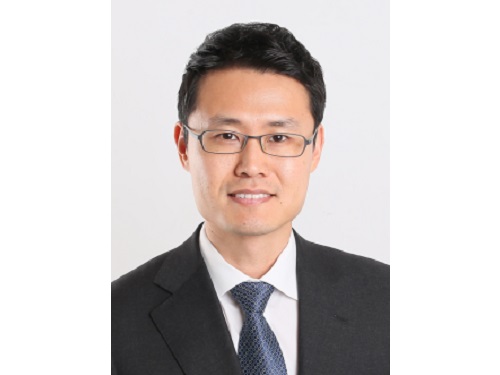
< Photo 1. (From left) KAIST ME Prof. Hae-Won Park, Ph.D. Student Yong Um, Ph.D. Student Seungwoo Hong >
- Professor Hae-Won Park's team at the Department of Mechanical Engineering developed a quadrupedal robot that can move at a high speed on ferrous walls and ceilings.
- It is expected to make a wide variety of contributions as it is to be used to conduct inspections and repairs of large steel structures such as ships, bridges, and transmission towers, offering an alternative to dangerous or risky activities required in hazardous environments while maintaining productivity and efficiency through automation and unmanning of such operations.
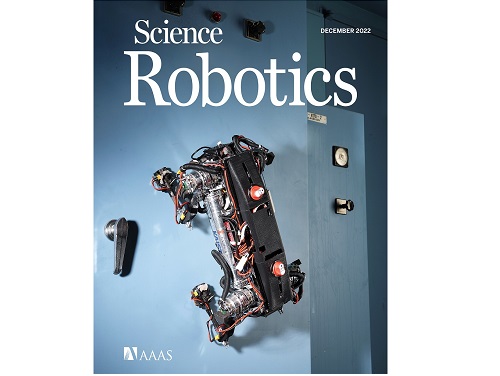
- The study was published as the cover paper of the December issue of Science Robotics.
KAIST (President Kwang Hyung Lee) announced on the 26th that a research team led by Professor Hae-Won Park of the Department of Mechanical Engineering developed a quadrupedal walking robot that can move at high speed on steel walls and ceilings named M.A.R.V.E.L. - rightly so as it is a Magnetically Adhesive Robot for Versatile and Expeditious Locomotion as described in their paper, “Agile and Versatile Climbing on Ferromagnetic Surfaces with a Quadrupedal Robot.” (DOI: 10.1126/scirobotics.add1017)
To make this happen, Professor Park's research team developed a foot pad that can quickly turn the magnetic adhesive force on and off while retaining high adhesive force even on an uneven surface through the use of the Electro-Permanent Magnet (EPM), a device that can magnetize and demagnetize an electromagnet with little power, and the Magneto-Rheological Elastomer (MRE), an elastic material made by mixing a magnetic response factor, such as iron powder, with an elastic material, such as rubber, which they mounted on a small quadrupedal robot they made in-house, at their own laboratory. These walking robots are expected to be put into a wide variety of usage, including being programmed to perform inspections, repairs, and maintenance tasks on large structures made of steel, such as ships, bridges, transmission towers, large storage areas, and construction sites.
This study, in which Seungwoo Hong and Yong Um of the Department of Mechanical Engineering participated as co-first authors, was published as the cover paper in the December issue of Science Robotics.
< Image on the Cover of 2022 December issue of Science Robotics >
Existing wall-climbing robots use wheels or endless tracks, so their mobility is limited on surfaces with steps or irregularities. On the other hand, walking robots for climbing can expect improved mobility in obstacle terrain, but have disadvantages in that they have significantly slower moving speeds or cannot perform various movements.
In order to enable fast movement of the walking robot, the sole of the foot must have strong adhesion force and be able to control the adhesion to quickly switch from sticking to the surface or to be off of it. In addition, it is necessary to maintain the adhesion force even on a rough or uneven surface.
To solve this problem, the research team used the EPM and MRE for the first time in designing the soles of walking robots. An EPM is a magnet that can turn on and off the electromagnetic force with a short current pulse. Unlike general electromagnets, it has the advantage that it does not require energy to maintain the magnetic force. The research team proposed a new EPM with a rectangular structure arrangement, enabling faster switching while significantly lowering the voltage required for switching compared to existing electromagnets.
In addition, the research team was able to increase the frictional force without significantly reducing the magnetic force of the sole by covering the sole with an MRE. The proposed sole weighs only 169 g, but provides a vertical gripping force of about *535 Newtons (N) and a frictional force of 445 N, which is sufficient gripping force for a quadrupedal robot weighing 8 kg.
* 535 N converted to kg is 54.5 kg, and 445 N is 45.4 kg. In other words, even if an external force of up to 54.5 kg in the vertical direction and up to 45.4 kg in the horizontal direction is applied (or even if a corresponding weight is hung), the sole of the foot does not come off the steel plate.
MARVEL climbed up a vertical wall at high speed at a speed of 70 cm per second, and was able to walk while hanging upside down from the ceiling at a maximum speed of 50 cm per second. This is the world's fastest speed for a walking climbing robot. In addition, the research team demonstrated that the robot can climb at a speed of up to 35 cm even on a surface that is painted, dirty with dust and the rust-tainted surfaces of water tanks, proving the robot's performance in a real environment. It was experimentally demonstrated that the robot not only exhibited high speed, but also can switch from floor to wall and from wall to ceiling, and overcome 5-cm high obstacles protruding from walls without difficulty.
The new climbing quadrupedal robot is expected to be widely used for inspection, repair, and maintenance of large steel structures such as ships, bridges, transmission towers, oil pipelines, large storage areas, and construction sites. As the works required in these places involves risks such as falls, suffocation and other accidents that may result in serious injuries or casualties, the need for automation is of utmost urgency.
One of the first co-authors of the paper, a Ph.D. student, Yong Um of KAIST’s Department of Mechanical Engineering, said, "By the use of the magnetic soles made up of the EPM and MRE and the non-linear model predictive controller suitable for climbing, the robot can speedily move through a variety of ferromagnetic surfaces including walls and ceilings, not just level grounds. We believe this would become a cornerstone that will expand the mobility and the places of pedal-mobile robots can venture into." He added, “These robots can be put into good use in executing dangerous and difficult tasks on steel structures in places like the shipbuilding yards.”
This research was carried out with support from the National Research Foundation of Korea's Basic Research in Science & Engineering Program for Mid-Career Researchers and Korea Shipbuilding & Offshore Engineering Co., Ltd..
< Figure 1. The quadrupedal robot (MARVEL) walking over various ferrous surfaces. (A) vertical wall (B) ceiling. (C) over obstacles on a vertical wall (D) making floor-to-wall and wall-to-ceiling transitions (E) moving over a storage tank (F) walking on a wall with a 2-kg weight and over a ceiling with a 3-kg load. >
< Figure 2. Description of the magnetic foot (A) Components of the magnet sole: ankle, Square Eletro-Permanent Magnet(S-EPM), MRE footpad. (B) Components of the S-EPM and MRE footpad. (C) Working principle of the S-EPM. When the magnetization direction is aligned as shown in the left figure, magnetic flux comes out of the keeper and circulates through the steel plate, generating holding force (ON state). Conversely, if the magnetization direction is aligned as shown in the figure on the right, the magnetic flux circulates inside the S-EPM and the holding force disappears (OFF state). >
Video Introduction: Agile and versatile climbing on ferromagnetic surfaces with a quadrupedal robot - YouTube
2022.12.30 View 20581
A Quick but Clingy Creepy-Crawler that will MARVEL You
Engineered by KAIST Mechanics, a quadrupedal robot climbs steel walls and crawls across metal ceilings at the fastest speed that the world has ever seen.
< Photo 1. (From left) KAIST ME Prof. Hae-Won Park, Ph.D. Student Yong Um, Ph.D. Student Seungwoo Hong >
- Professor Hae-Won Park's team at the Department of Mechanical Engineering developed a quadrupedal robot that can move at a high speed on ferrous walls and ceilings.
- It is expected to make a wide variety of contributions as it is to be used to conduct inspections and repairs of large steel structures such as ships, bridges, and transmission towers, offering an alternative to dangerous or risky activities required in hazardous environments while maintaining productivity and efficiency through automation and unmanning of such operations.
- The study was published as the cover paper of the December issue of Science Robotics.
KAIST (President Kwang Hyung Lee) announced on the 26th that a research team led by Professor Hae-Won Park of the Department of Mechanical Engineering developed a quadrupedal walking robot that can move at high speed on steel walls and ceilings named M.A.R.V.E.L. - rightly so as it is a Magnetically Adhesive Robot for Versatile and Expeditious Locomotion as described in their paper, “Agile and Versatile Climbing on Ferromagnetic Surfaces with a Quadrupedal Robot.” (DOI: 10.1126/scirobotics.add1017)
To make this happen, Professor Park's research team developed a foot pad that can quickly turn the magnetic adhesive force on and off while retaining high adhesive force even on an uneven surface through the use of the Electro-Permanent Magnet (EPM), a device that can magnetize and demagnetize an electromagnet with little power, and the Magneto-Rheological Elastomer (MRE), an elastic material made by mixing a magnetic response factor, such as iron powder, with an elastic material, such as rubber, which they mounted on a small quadrupedal robot they made in-house, at their own laboratory. These walking robots are expected to be put into a wide variety of usage, including being programmed to perform inspections, repairs, and maintenance tasks on large structures made of steel, such as ships, bridges, transmission towers, large storage areas, and construction sites.
This study, in which Seungwoo Hong and Yong Um of the Department of Mechanical Engineering participated as co-first authors, was published as the cover paper in the December issue of Science Robotics.
< Image on the Cover of 2022 December issue of Science Robotics >
Existing wall-climbing robots use wheels or endless tracks, so their mobility is limited on surfaces with steps or irregularities. On the other hand, walking robots for climbing can expect improved mobility in obstacle terrain, but have disadvantages in that they have significantly slower moving speeds or cannot perform various movements.
In order to enable fast movement of the walking robot, the sole of the foot must have strong adhesion force and be able to control the adhesion to quickly switch from sticking to the surface or to be off of it. In addition, it is necessary to maintain the adhesion force even on a rough or uneven surface.
To solve this problem, the research team used the EPM and MRE for the first time in designing the soles of walking robots. An EPM is a magnet that can turn on and off the electromagnetic force with a short current pulse. Unlike general electromagnets, it has the advantage that it does not require energy to maintain the magnetic force. The research team proposed a new EPM with a rectangular structure arrangement, enabling faster switching while significantly lowering the voltage required for switching compared to existing electromagnets.
In addition, the research team was able to increase the frictional force without significantly reducing the magnetic force of the sole by covering the sole with an MRE. The proposed sole weighs only 169 g, but provides a vertical gripping force of about *535 Newtons (N) and a frictional force of 445 N, which is sufficient gripping force for a quadrupedal robot weighing 8 kg.
* 535 N converted to kg is 54.5 kg, and 445 N is 45.4 kg. In other words, even if an external force of up to 54.5 kg in the vertical direction and up to 45.4 kg in the horizontal direction is applied (or even if a corresponding weight is hung), the sole of the foot does not come off the steel plate.
MARVEL climbed up a vertical wall at high speed at a speed of 70 cm per second, and was able to walk while hanging upside down from the ceiling at a maximum speed of 50 cm per second. This is the world's fastest speed for a walking climbing robot. In addition, the research team demonstrated that the robot can climb at a speed of up to 35 cm even on a surface that is painted, dirty with dust and the rust-tainted surfaces of water tanks, proving the robot's performance in a real environment. It was experimentally demonstrated that the robot not only exhibited high speed, but also can switch from floor to wall and from wall to ceiling, and overcome 5-cm high obstacles protruding from walls without difficulty.
The new climbing quadrupedal robot is expected to be widely used for inspection, repair, and maintenance of large steel structures such as ships, bridges, transmission towers, oil pipelines, large storage areas, and construction sites. As the works required in these places involves risks such as falls, suffocation and other accidents that may result in serious injuries or casualties, the need for automation is of utmost urgency.
One of the first co-authors of the paper, a Ph.D. student, Yong Um of KAIST’s Department of Mechanical Engineering, said, "By the use of the magnetic soles made up of the EPM and MRE and the non-linear model predictive controller suitable for climbing, the robot can speedily move through a variety of ferromagnetic surfaces including walls and ceilings, not just level grounds. We believe this would become a cornerstone that will expand the mobility and the places of pedal-mobile robots can venture into." He added, “These robots can be put into good use in executing dangerous and difficult tasks on steel structures in places like the shipbuilding yards.”
This research was carried out with support from the National Research Foundation of Korea's Basic Research in Science & Engineering Program for Mid-Career Researchers and Korea Shipbuilding & Offshore Engineering Co., Ltd..
< Figure 1. The quadrupedal robot (MARVEL) walking over various ferrous surfaces. (A) vertical wall (B) ceiling. (C) over obstacles on a vertical wall (D) making floor-to-wall and wall-to-ceiling transitions (E) moving over a storage tank (F) walking on a wall with a 2-kg weight and over a ceiling with a 3-kg load. >
< Figure 2. Description of the magnetic foot (A) Components of the magnet sole: ankle, Square Eletro-Permanent Magnet(S-EPM), MRE footpad. (B) Components of the S-EPM and MRE footpad. (C) Working principle of the S-EPM. When the magnetization direction is aligned as shown in the left figure, magnetic flux comes out of the keeper and circulates through the steel plate, generating holding force (ON state). Conversely, if the magnetization direction is aligned as shown in the figure on the right, the magnetic flux circulates inside the S-EPM and the holding force disappears (OFF state). >
Video Introduction: Agile and versatile climbing on ferromagnetic surfaces with a quadrupedal robot - YouTube
2022.12.30 View 20581 -
 KAIST Offers Hope to Musicians with Dystonia
< Photo 1. Conductor and Pianist João Carlos Martins before the Recital at the Carnegie Hall preparing with his bionic gloves >
KAIST’s neuroscientist and professor, Dr. Daesoo Kim attended the “Conference for Musicians with Dystonia” supported by the World Health Organization (WHO) and the Carnegie Hall concert of legendary pianist João Carlos Martins, who is also a dystonia patient, to announce his team’s recent advancements toward finding a cure for dystonia.
On November 19, 2022, a “miracle concert” was held in Carnegie Hall. João Carlos Martins was a renowned world-class pianist in the 70s and 80s, but he had to put an end to his musical career due to focal dystonia in his fingers. But in 2020, he began using a bionic glove developed by industrial designer Ubiratã Bizarro Costa and after years of hard work he was back in Carnegie Hall as an 82-year-old man.
During the concert, he conducted the NOVUS NY orchestra in a performance of Bach, and later even played the piano himself. In particular, between his performances, he gave shout-outs to scientists studying dystonia including KAIST Professor Daesoo Kim, asking them to continue working towards curing rare diseases for musicians.
< Photo 2. Professor Daesoo Kim with Conductor and Pianist João Carlos Martins >
Musician’s dystonia affects 1-3% of musicians around the world and musicians make up approximately 5% of the total number of dystonia patients. Musicians who are no longer able to practice music due to the disease often experience stress and depression, which may even lead to suicide in extreme cases. Musicians are known to be particularly prone to such diseases due to excessive practice regimens, perfectionism, and even genetics. Currently, botulinum toxin (Botox) is used to suppress abnormal muscles, but muscle function suppression ultimately means that the musician is no longer able to play the instrument. João Carlos Martins himself underwent several Botox procedures and three brain surgeries, but saw no therapeutic results. This is why a new treatment was necessary.
Professor Daesoo Kim’s research team at KAIST took note of the fact that abnormal muscle tension is caused by excessive stress, and developed NT-1, a treatment that blocks the development of the symptoms of dystonia from the brain, allowing patients to use their muscles as they normally would. The research team published their findings in Science Advances in 2021, and João Carlos Martins invited Professor Daesoo Kim to the UN conference and his concert after reading this paper.
< Photo 3. Professor Daesoo Kim (3rd from the left) photographed with other guests at the recital including Dr. Dévora Kestel, the Director of the Mental Health and Substance Use at WHO, sharing the center with Conductor and Pianist João Carlos Martins >
During the UN conference held the day prior to the Carnegie Hall concert, Dr. Dévora Kestel, Director of the Mental Health and Substance Use at WHO, said, “Although dystonia is not as well-known, it is a common disease around the world, and needs our society’s attention and the devotion of many researchers.” Professor Daesoo Kim said, “NT-1 is a drug that blocks the cause of dystonia in the brain, and will allow musicians to continue practicing music. We aim to attain clinical approval in Korea by 2024.”
NT-1 is currently under development by NeuroTobe, a faculty-led start-up company at KAIST, headed by Professor Daesoo Kim as the CEO. The synthesis of the drug for clinical testing has been successfully completed, and it has shown excellent efficacy and safety through various rounds of animal testing. Unlike Botox, which takes a few days to show its therapeutic effects after receiving the procedure from a hospital, NT-1 shows its therapeutic effects within an hour after taking it. As a so-called “edible Botox”, it is expected to help treat various muscular diseases and ailments.
2022.12.27 View 13455
KAIST Offers Hope to Musicians with Dystonia
< Photo 1. Conductor and Pianist João Carlos Martins before the Recital at the Carnegie Hall preparing with his bionic gloves >
KAIST’s neuroscientist and professor, Dr. Daesoo Kim attended the “Conference for Musicians with Dystonia” supported by the World Health Organization (WHO) and the Carnegie Hall concert of legendary pianist João Carlos Martins, who is also a dystonia patient, to announce his team’s recent advancements toward finding a cure for dystonia.
On November 19, 2022, a “miracle concert” was held in Carnegie Hall. João Carlos Martins was a renowned world-class pianist in the 70s and 80s, but he had to put an end to his musical career due to focal dystonia in his fingers. But in 2020, he began using a bionic glove developed by industrial designer Ubiratã Bizarro Costa and after years of hard work he was back in Carnegie Hall as an 82-year-old man.
During the concert, he conducted the NOVUS NY orchestra in a performance of Bach, and later even played the piano himself. In particular, between his performances, he gave shout-outs to scientists studying dystonia including KAIST Professor Daesoo Kim, asking them to continue working towards curing rare diseases for musicians.
< Photo 2. Professor Daesoo Kim with Conductor and Pianist João Carlos Martins >
Musician’s dystonia affects 1-3% of musicians around the world and musicians make up approximately 5% of the total number of dystonia patients. Musicians who are no longer able to practice music due to the disease often experience stress and depression, which may even lead to suicide in extreme cases. Musicians are known to be particularly prone to such diseases due to excessive practice regimens, perfectionism, and even genetics. Currently, botulinum toxin (Botox) is used to suppress abnormal muscles, but muscle function suppression ultimately means that the musician is no longer able to play the instrument. João Carlos Martins himself underwent several Botox procedures and three brain surgeries, but saw no therapeutic results. This is why a new treatment was necessary.
Professor Daesoo Kim’s research team at KAIST took note of the fact that abnormal muscle tension is caused by excessive stress, and developed NT-1, a treatment that blocks the development of the symptoms of dystonia from the brain, allowing patients to use their muscles as they normally would. The research team published their findings in Science Advances in 2021, and João Carlos Martins invited Professor Daesoo Kim to the UN conference and his concert after reading this paper.
< Photo 3. Professor Daesoo Kim (3rd from the left) photographed with other guests at the recital including Dr. Dévora Kestel, the Director of the Mental Health and Substance Use at WHO, sharing the center with Conductor and Pianist João Carlos Martins >
During the UN conference held the day prior to the Carnegie Hall concert, Dr. Dévora Kestel, Director of the Mental Health and Substance Use at WHO, said, “Although dystonia is not as well-known, it is a common disease around the world, and needs our society’s attention and the devotion of many researchers.” Professor Daesoo Kim said, “NT-1 is a drug that blocks the cause of dystonia in the brain, and will allow musicians to continue practicing music. We aim to attain clinical approval in Korea by 2024.”
NT-1 is currently under development by NeuroTobe, a faculty-led start-up company at KAIST, headed by Professor Daesoo Kim as the CEO. The synthesis of the drug for clinical testing has been successfully completed, and it has shown excellent efficacy and safety through various rounds of animal testing. Unlike Botox, which takes a few days to show its therapeutic effects after receiving the procedure from a hospital, NT-1 shows its therapeutic effects within an hour after taking it. As a so-called “edible Botox”, it is expected to help treat various muscular diseases and ailments.
2022.12.27 View 13455 -
 KAIST Team Develops Surface-Lighting MicroLED Patch with Significant Melanogenesis Inhibition Effect
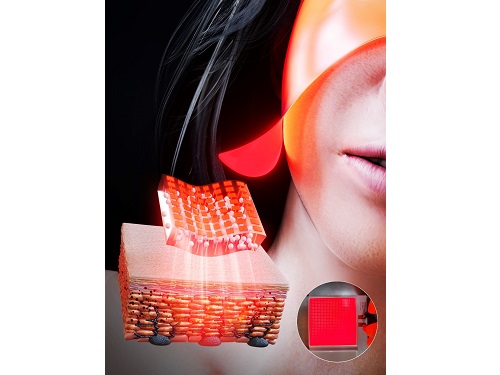
A KAIST research team led by Ph.d candidate Jae Hee Lee and Professor Keon Jae Lee from the Department of Materials Science and Engineering has developed a surface-lighting microLED patch for UV-induced melanogenesis inhibition.
Melanin is brown or dark pigments existing in the skin, which can be abnormally synthesized by external UV or stress. Since the excessive melanin leads to skin diseases such as spots and freckles, proper treatment is required to return normal skin condition.
Recently, LED-based photo-stimulators have been released for skin care, however, their therapeutic effect is still controversial. Since conventional LED stimulators cannot conformally attach to the human skin, distance-induced side effects are caused by light loss and high heat transfer. To achieve effective phototreatment, the LED stimulator needs to be irradiated in contact with the human skin surface, enabling proper and uniform light deliver to the dermis with minimal optical loss.
In this work, the research team fabricated skin-attachable surface-lighting microLED (SµLED, 4 × 4 cm2) patch by utilizing a thousand of microLED chips and silica-embedded light diffusion layer. 100 µm-sized LED chips are vertically-interconnected for high flexibility and low heat generation, allowing its long-term operation on the human skin.
< Image 1. The overall concept of SµLED patch. a) SµLED patch operated on the human skin. b) Schematic illustration of SµLED patch structure. c) 4 × 4 cm2-sized SµLED patch. d) Schematic illustration of the advantages of SµLED patch such as efficient light delivery, low heat generation, and surface-lighting irradiation. >
The research team confirmed melanogenesis inhibition by irradiating the SµLED patch and the conventional LED (CLED) on the artificial human skin and mice dorsal skin. The SµLED-treated groups of human cells and mouse tissues showed minimal epidermal photo-toxicity and consistently effective reduction in synthesized melanin, compared to CLED-treated groups. In addition, significant suppression of proteins/catalysts expression involved in melanin synthesis such as MITF (microphthalmia-associated transcription factor), Melan-A and tyrosinase was verified.
< Image 2. The efficacy of melanogenesis inhibition on 3D human skin cells. a). Different irradiation conditions for a-MSH (major factor to stimulate melanin synthesis) treated cells. b) The ratio of pigmented area to total epidermis area. c) Relative variance of melanin level in 1 cm2-sized skin cells. A low variance means that melanin is evenly distributed, and a high variance means that the melanin is irregularly distributed. d) Optical images after in vitro experiments for 12 days. Scale bar, 1cm. e) Histological analysis of 3D skin, showing the greatest reduction in melanin after SµLED irradiation. Scale bar, 20 µm. >
< Image 3. The efficacy of melanogenesis inhibition on mouse dorsal skin. a) Optical images of mice dorsal skin after photo-treatment for 20 days. b) Histological analysis of mice dorsal skin. Less brown color means less expression of protein/catalysis involved in melanin synthesis. Scale bar, 50 µm. >
Prof. Keon Jae Lee said, “Our inorganic-based SµLED patch has outstanding characteristics in light efficiency, reliability, and durability. The SµLED patch is expected to give a great impact on the cosmetic field by reducing side effects and maximizing phototherapeutic effects.” The core technology of cosmetic SµLED has been transferred to Fronics co., Ltd, founded by Prof. Lee. Fronics is building foundry and equipment for mass production of SµLED masks for whole face cover and plans to release the products in March next year.
This paper entitled “Wearable Surface-Lighting Micro-Light-Emitting Diode Patch for Melanogenesis Inhibition” was published in the November 2022 issue of Advanced Healthcare Materials.
2022.11.22 View 14576
KAIST Team Develops Surface-Lighting MicroLED Patch with Significant Melanogenesis Inhibition Effect
A KAIST research team led by Ph.d candidate Jae Hee Lee and Professor Keon Jae Lee from the Department of Materials Science and Engineering has developed a surface-lighting microLED patch for UV-induced melanogenesis inhibition.
Melanin is brown or dark pigments existing in the skin, which can be abnormally synthesized by external UV or stress. Since the excessive melanin leads to skin diseases such as spots and freckles, proper treatment is required to return normal skin condition.
Recently, LED-based photo-stimulators have been released for skin care, however, their therapeutic effect is still controversial. Since conventional LED stimulators cannot conformally attach to the human skin, distance-induced side effects are caused by light loss and high heat transfer. To achieve effective phototreatment, the LED stimulator needs to be irradiated in contact with the human skin surface, enabling proper and uniform light deliver to the dermis with minimal optical loss.
In this work, the research team fabricated skin-attachable surface-lighting microLED (SµLED, 4 × 4 cm2) patch by utilizing a thousand of microLED chips and silica-embedded light diffusion layer. 100 µm-sized LED chips are vertically-interconnected for high flexibility and low heat generation, allowing its long-term operation on the human skin.
< Image 1. The overall concept of SµLED patch. a) SµLED patch operated on the human skin. b) Schematic illustration of SµLED patch structure. c) 4 × 4 cm2-sized SµLED patch. d) Schematic illustration of the advantages of SµLED patch such as efficient light delivery, low heat generation, and surface-lighting irradiation. >
The research team confirmed melanogenesis inhibition by irradiating the SµLED patch and the conventional LED (CLED) on the artificial human skin and mice dorsal skin. The SµLED-treated groups of human cells and mouse tissues showed minimal epidermal photo-toxicity and consistently effective reduction in synthesized melanin, compared to CLED-treated groups. In addition, significant suppression of proteins/catalysts expression involved in melanin synthesis such as MITF (microphthalmia-associated transcription factor), Melan-A and tyrosinase was verified.
< Image 2. The efficacy of melanogenesis inhibition on 3D human skin cells. a). Different irradiation conditions for a-MSH (major factor to stimulate melanin synthesis) treated cells. b) The ratio of pigmented area to total epidermis area. c) Relative variance of melanin level in 1 cm2-sized skin cells. A low variance means that melanin is evenly distributed, and a high variance means that the melanin is irregularly distributed. d) Optical images after in vitro experiments for 12 days. Scale bar, 1cm. e) Histological analysis of 3D skin, showing the greatest reduction in melanin after SµLED irradiation. Scale bar, 20 µm. >
< Image 3. The efficacy of melanogenesis inhibition on mouse dorsal skin. a) Optical images of mice dorsal skin after photo-treatment for 20 days. b) Histological analysis of mice dorsal skin. Less brown color means less expression of protein/catalysis involved in melanin synthesis. Scale bar, 50 µm. >
Prof. Keon Jae Lee said, “Our inorganic-based SµLED patch has outstanding characteristics in light efficiency, reliability, and durability. The SµLED patch is expected to give a great impact on the cosmetic field by reducing side effects and maximizing phototherapeutic effects.” The core technology of cosmetic SµLED has been transferred to Fronics co., Ltd, founded by Prof. Lee. Fronics is building foundry and equipment for mass production of SµLED masks for whole face cover and plans to release the products in March next year.
This paper entitled “Wearable Surface-Lighting Micro-Light-Emitting Diode Patch for Melanogenesis Inhibition” was published in the November 2022 issue of Advanced Healthcare Materials.
2022.11.22 View 14576 -
 Phage resistant Escherichia coli strains developed to reduce fermentation failure
A genome engineering-based systematic strategy for developing phage resistant Escherichia coli strains has been successfully developed through the collaborative efforts of a team led by Professor Sang Yup Lee, Professor Shi Chen, and Professor Lianrong Wang. This study by Xuan Zou et al. was published in Nature Communications in August 2022 and featured in Nature Communications Editors’ Highlights. The collaboration by the School of Pharmaceutical Sciences at Wuhan University, the First Affiliated Hospital of Shenzhen University, and the KAIST Department of Chemical and Biomolecular Engineering has made an important advance in the metabolic engineering and fermentation industry as it solves a big problem of phage infection causing fermentation failure.
Systems metabolic engineering is a highly interdisciplinary field that has made the development of microbial cell factories to produce various bioproducts including chemicals, fuels, and materials possible in a sustainable and environmentally friendly way, mitigating the impact of worldwide resource depletion and climate change. Escherichia coli is one of the most important chassis microbial strains, given its wide applications in the bio-based production of a diverse range of chemicals and materials. With the development of tools and strategies for systems metabolic engineering using E. coli, a highly optimized and well-characterized cell factory will play a crucial role in converting cheap and readily available raw materials into products of great economic and industrial value.
However, the consistent problem of phage contamination in fermentation imposes a devastating impact on host cells and threatens the productivity of bacterial bioprocesses in biotechnology facilities, which can lead to widespread fermentation failure and immeasurable economic loss. Host-controlled defense systems can be developed into effective genetic engineering solutions to address bacteriophage contamination in industrial-scale fermentation; however, most of the resistance mechanisms only narrowly restrict phages and their effect on phage contamination will be limited.
There have been attempts to develop diverse abilities/systems for environmental adaptation or antiviral defense. The team’s collaborative efforts developed a new type II single-stranded DNA phosphorothioation (Ssp) defense system derived from E. coli 3234/A, which can be used in multiple industrial E. coli strains (e.g., E. coli K-12, B and W) to provide broad protection against various types of dsDNA coliphages. Furthermore, they developed a systematic genome engineering strategy involving the simultaneous genomic integration of the Ssp defense module and mutations in components that are essential to the phage life cycle. This strategy can be used to transform E. coli hosts that are highly susceptible to phage attack into strains with powerful restriction effects on the tested bacteriophages. This endows hosts with strong resistance against a wide spectrum of phage infections without affecting bacterial growth and normal physiological function. More importantly, the resulting engineered phage-resistant strains maintained the capabilities of producing the desired chemicals and recombinant proteins even under high levels of phage cocktail challenge, which provides crucial protection against phage attacks.
This is a major step forward, as it provides a systematic solution for engineering phage-resistant bacterial strains, especially industrial bioproduction strains, to protect cells from a wide range of bacteriophages. Considering the functionality of this engineering strategy with diverse E. coli strains, the strategy reported in this study can be widely extended to other bacterial species and industrial applications, which will be of great interest to researchers in academia and industry alike.
Fig. A schematic model of the systematic strategy for engineering phage-sensitive industrial E. coli strains into strains with broad antiphage activities. Through the simultaneous genomic integration of a DNA phosphorothioation-based Ssp defense module and mutations of components essential for the phage life cycle, the engineered E. coli strains show strong resistance against diverse phages tested and maintain the capabilities of producing example recombinant proteins, even under high levels of phage cocktail challenge.
2022.08.23 View 17125
Phage resistant Escherichia coli strains developed to reduce fermentation failure
A genome engineering-based systematic strategy for developing phage resistant Escherichia coli strains has been successfully developed through the collaborative efforts of a team led by Professor Sang Yup Lee, Professor Shi Chen, and Professor Lianrong Wang. This study by Xuan Zou et al. was published in Nature Communications in August 2022 and featured in Nature Communications Editors’ Highlights. The collaboration by the School of Pharmaceutical Sciences at Wuhan University, the First Affiliated Hospital of Shenzhen University, and the KAIST Department of Chemical and Biomolecular Engineering has made an important advance in the metabolic engineering and fermentation industry as it solves a big problem of phage infection causing fermentation failure.
Systems metabolic engineering is a highly interdisciplinary field that has made the development of microbial cell factories to produce various bioproducts including chemicals, fuels, and materials possible in a sustainable and environmentally friendly way, mitigating the impact of worldwide resource depletion and climate change. Escherichia coli is one of the most important chassis microbial strains, given its wide applications in the bio-based production of a diverse range of chemicals and materials. With the development of tools and strategies for systems metabolic engineering using E. coli, a highly optimized and well-characterized cell factory will play a crucial role in converting cheap and readily available raw materials into products of great economic and industrial value.
However, the consistent problem of phage contamination in fermentation imposes a devastating impact on host cells and threatens the productivity of bacterial bioprocesses in biotechnology facilities, which can lead to widespread fermentation failure and immeasurable economic loss. Host-controlled defense systems can be developed into effective genetic engineering solutions to address bacteriophage contamination in industrial-scale fermentation; however, most of the resistance mechanisms only narrowly restrict phages and their effect on phage contamination will be limited.
There have been attempts to develop diverse abilities/systems for environmental adaptation or antiviral defense. The team’s collaborative efforts developed a new type II single-stranded DNA phosphorothioation (Ssp) defense system derived from E. coli 3234/A, which can be used in multiple industrial E. coli strains (e.g., E. coli K-12, B and W) to provide broad protection against various types of dsDNA coliphages. Furthermore, they developed a systematic genome engineering strategy involving the simultaneous genomic integration of the Ssp defense module and mutations in components that are essential to the phage life cycle. This strategy can be used to transform E. coli hosts that are highly susceptible to phage attack into strains with powerful restriction effects on the tested bacteriophages. This endows hosts with strong resistance against a wide spectrum of phage infections without affecting bacterial growth and normal physiological function. More importantly, the resulting engineered phage-resistant strains maintained the capabilities of producing the desired chemicals and recombinant proteins even under high levels of phage cocktail challenge, which provides crucial protection against phage attacks.
This is a major step forward, as it provides a systematic solution for engineering phage-resistant bacterial strains, especially industrial bioproduction strains, to protect cells from a wide range of bacteriophages. Considering the functionality of this engineering strategy with diverse E. coli strains, the strategy reported in this study can be widely extended to other bacterial species and industrial applications, which will be of great interest to researchers in academia and industry alike.
Fig. A schematic model of the systematic strategy for engineering phage-sensitive industrial E. coli strains into strains with broad antiphage activities. Through the simultaneous genomic integration of a DNA phosphorothioation-based Ssp defense module and mutations of components essential for the phage life cycle, the engineered E. coli strains show strong resistance against diverse phages tested and maintain the capabilities of producing example recombinant proteins, even under high levels of phage cocktail challenge.
2022.08.23 View 17125 -
 KAIST Research Team Proves How a Neurotransmitter may be the Key in Controlling Alzheimer’s Toxicity
With nearly 50 million dementia patients worldwide, and Alzheimers’s disease is the most common neurodegenerative disease. Its main symptom is the impairment of general cognitive abilities, including the ability to speak or to remember. The importance of finding a cure is widely understood with increasingly aging population and the life expectancy being ever-extended. However, even the cause of the grim disease is yet to be given a clear definition.
A KAIST research team in the Department of Chemistry led by professor Mi Hee Lim took on a lead to discovered a new role for somatostatin, a protein-based neurotransmitter, in reducing the toxicity caused in the pathogenic mechanism taken towards development of Alzheimer’s disease. The study was published in the July issue of Nature Chemistry under the title, “Conformational and functional changes of the native neuropeptide somatostatin occur in the presence of copper and amyloid-β”.
According to the amyloid hypothesis, the abnormal deposition of Aβ proteins causes death of neuronal cells. While Aβ agglomerations make up most of the aged plaques through fibrosis, in recent studies, high concentrations of transitional metal were found in the plaques from Alzheimer’s patients.
This suggests a close interaction between metallic ions and Aβ, which accelerates the fibrosis of proteins. Copper in particular is a redox-activating transition metal that can produce large amounts of oxygen and cause serious oxidative stress on cell organelles. Aβ proteins and transition metals can closely interact with neurotransmitters at synapses, but the direct effects of such abnormalities on the structure and function of neurotransmitters are yet to be understood.
Figure 1. Functional shift of somatostatin (SST) by factors in the pathogenesis of Alzheimer's disease.
Figure 2. Somatostatin’s loss-of-function as neurotransmitter. a. Schematic diagram of SST auto-aggregation due to Alzheimer's pathological factors. b. SST’s aggregation by copper ions. c. Coordination-prediction structure and N-terminal folding of copper-SST. d. Inhibition of SST receptor binding specificity by metals.
In their research, Professor Lim’s team discovered that when somatostatin, the protein-based neurotransmitter, is met with copper, Aβ, and metal-Aβ complexes, self-aggregates and ceases to perform its innate function of transmitting neural signals, but begins to attenuate the toxicity and agglomeration of metal-Aβ complexes.
Figure 3. Gain-of-function of somatostatin (SST) in the dementia setting. a. Prediction of docking of SST and amyloid beta. b. SST making metal-amyloid beta aggregates into an amorphous form. c. Cytotoxic mitigation effect of SST. d. SST mitigating the interaction between amyloid beta protein with the cell membrane.
This research, by Dr. Jiyeon Han et al. from the KAIST Department of Chemistry, revealed the coordination structure between copper and somatostatin at a molecular level through which it suggested the agglomeration mechanism, and discovered the effects of somatostatin on Aβ agglomeration path depending on the presence or absence of metals. The team has further confirmed somatostatin’s receptor binding, interactions with cell membranes, and effects on cell toxicity for the first time to receive international attention.
Professor Mi Hee Lim said, “This research has great significance in having discovered a new role of neurotransmitters in the pathogenesis of Alzheimer’s disease.” “We expect this research to contribute to defining the pathogenic network of neurodegenerative diseases caused by aging, and to the development of future biomarkers and medicine,” she added.
This research was conducted jointly by Professor Seung-Hee Lee’s team of KAIST Department of Biological Sciences, Professor Kiyoung Park’s Team of KAIST Department of Chemistry, and Professor Yulong Li’s team of Peking University.
The research was funded by Basic Science Research Program of the National Research Foundation of Korea and KAIST.
For more information about the research team, visit the website: https://sites.google.com/site/miheelimlab/1-professor-mi-hee-lim.
2022.07.29 View 17522
KAIST Research Team Proves How a Neurotransmitter may be the Key in Controlling Alzheimer’s Toxicity
With nearly 50 million dementia patients worldwide, and Alzheimers’s disease is the most common neurodegenerative disease. Its main symptom is the impairment of general cognitive abilities, including the ability to speak or to remember. The importance of finding a cure is widely understood with increasingly aging population and the life expectancy being ever-extended. However, even the cause of the grim disease is yet to be given a clear definition.
A KAIST research team in the Department of Chemistry led by professor Mi Hee Lim took on a lead to discovered a new role for somatostatin, a protein-based neurotransmitter, in reducing the toxicity caused in the pathogenic mechanism taken towards development of Alzheimer’s disease. The study was published in the July issue of Nature Chemistry under the title, “Conformational and functional changes of the native neuropeptide somatostatin occur in the presence of copper and amyloid-β”.
According to the amyloid hypothesis, the abnormal deposition of Aβ proteins causes death of neuronal cells. While Aβ agglomerations make up most of the aged plaques through fibrosis, in recent studies, high concentrations of transitional metal were found in the plaques from Alzheimer’s patients.
This suggests a close interaction between metallic ions and Aβ, which accelerates the fibrosis of proteins. Copper in particular is a redox-activating transition metal that can produce large amounts of oxygen and cause serious oxidative stress on cell organelles. Aβ proteins and transition metals can closely interact with neurotransmitters at synapses, but the direct effects of such abnormalities on the structure and function of neurotransmitters are yet to be understood.
Figure 1. Functional shift of somatostatin (SST) by factors in the pathogenesis of Alzheimer's disease.
Figure 2. Somatostatin’s loss-of-function as neurotransmitter. a. Schematic diagram of SST auto-aggregation due to Alzheimer's pathological factors. b. SST’s aggregation by copper ions. c. Coordination-prediction structure and N-terminal folding of copper-SST. d. Inhibition of SST receptor binding specificity by metals.
In their research, Professor Lim’s team discovered that when somatostatin, the protein-based neurotransmitter, is met with copper, Aβ, and metal-Aβ complexes, self-aggregates and ceases to perform its innate function of transmitting neural signals, but begins to attenuate the toxicity and agglomeration of metal-Aβ complexes.
Figure 3. Gain-of-function of somatostatin (SST) in the dementia setting. a. Prediction of docking of SST and amyloid beta. b. SST making metal-amyloid beta aggregates into an amorphous form. c. Cytotoxic mitigation effect of SST. d. SST mitigating the interaction between amyloid beta protein with the cell membrane.
This research, by Dr. Jiyeon Han et al. from the KAIST Department of Chemistry, revealed the coordination structure between copper and somatostatin at a molecular level through which it suggested the agglomeration mechanism, and discovered the effects of somatostatin on Aβ agglomeration path depending on the presence or absence of metals. The team has further confirmed somatostatin’s receptor binding, interactions with cell membranes, and effects on cell toxicity for the first time to receive international attention.
Professor Mi Hee Lim said, “This research has great significance in having discovered a new role of neurotransmitters in the pathogenesis of Alzheimer’s disease.” “We expect this research to contribute to defining the pathogenic network of neurodegenerative diseases caused by aging, and to the development of future biomarkers and medicine,” she added.
This research was conducted jointly by Professor Seung-Hee Lee’s team of KAIST Department of Biological Sciences, Professor Kiyoung Park’s Team of KAIST Department of Chemistry, and Professor Yulong Li’s team of Peking University.
The research was funded by Basic Science Research Program of the National Research Foundation of Korea and KAIST.
For more information about the research team, visit the website: https://sites.google.com/site/miheelimlab/1-professor-mi-hee-lim.
2022.07.29 View 17522 -
 A System for Stable Simultaneous Communication among Thousands of IoT Devices
A mmWave Backscatter System, developed by a team led by Professor Song Min Kim is exciting news for the IoT market as it will be able to provide fast and stable connectivity even for a massive network, which could finally allow IoT devices to reach their full potential.
A research team led by Professor Song Min Kim of the KAIST School of Electrical Engineering developed a system that can support concurrent communications for tens of millions of IoT devices using backscattering millimeter-level waves (mmWave).
With their mmWave backscatter method, the research team built a design enabling simultaneous signal demodulation in a complex environment for communication where tens of thousands of IoT devices are arranged indoors. The wide frequency range of mmWave exceeds 10GHz, which provides great scalability. In addition, backscattering reflects radiated signals instead of wirelessly creating its own, which allows operation at ultralow power. Therefore, the mmWave backscatter system offers internet connectivity on a mass scale to IoT devices at a low installation cost.
This research by Kangmin Bae et al. was presented at ACM MobiSys 2022. At this world-renowned conference for mobile systems, the research won the Best Paper Award under the title “OmniScatter: Sensitivity mmWave Backscattering Using Commodity FMCW Radar”. It is meaningful that members of the KAIST School of Electrical Engineering have won the Best Paper Award at ACM MobiSys for two consecutive years, as last year was the first time the award was presented to an institute from Asia.
IoT, as a core component of 5G/6G network, is showing exponential growth, and is expected to be part of a trillion devices by 2035. To support the connection of IoT devices on a mass scale, 5G and 6G each aim to support ten times and 100 times the network density of 4G, respectively. As a result, the importance of practical systems for large-scale communication has been raised.
The mmWave is a next-generation communication technology that can be incorporated in 5G/6G standards, as it utilizes carrier waves at frequencies between 30 to 300GHz. However, due to signal reduction at high frequencies and reflection loss, the current mmWave backscatter system enables communication in limited environments. In other words, it cannot operate in complex environments where various obstacles and reflectors are present. As a result, it is limited to the large-scale connection of IoT devices that require a relatively free arrangement.
The research team found the solution in the high coding gain of an FMCW radar. The team developed a signal processing method that can fundamentally separate backscatter signals from ambient noise while maintaining the coding gain of the radar. They achieved a receiver sensitivity of over 100 thousand times that of previously reported FMCW radars, which can support communication in practical environments. Additionally, given the radar’s property where the frequency of the demodulated signal changes depending on the physical location of the tag, the team designed a system that passively assigns them channels. This lets the ultralow-power backscatter communication system to take full advantage of the frequency range at 10 GHz or higher.
The developed system can use the radar of existing commercial products as gateway, making it easily compatible. In addition, since the backscatter system works at ultralow power levels of 10uW or below, it can operate for over 40 years with a single button cell and drastically reduce installation and maintenance costs.
The research team confirmed that mmWave backscatter devices arranged randomly in an office with various obstacles and reflectors could communicate effectively. The team then took things one step further and conducted a successful trace-driven evaluation where they simultaneously received information sent by 1,100 devices.
Their research presents connectivity that greatly exceeds network density required by next-generation communication like 5G and 6G. The system is expected to become a stepping stone for the hyper-connected future to come.
Professor Kim said, “mmWave backscatter is the technology we’ve dreamt of. The mass scalability and ultralow power at which it can operate IoT devices is unmatched by any existing technology”. He added, “We look forward to this system being actively utilized to enable the wide availability of IoT in the hyper-connected generation to come”.
To demonstrate the massive connectivity of the system, a trace-driven evaluation of 1,100 concurrent tag transmissions are made. Figure shows the demodulation result of each and every 1,100 tags as red triangles, where they successfully communicate without collision.
This work was supported by Samsung Research Funding & Incubation Center of Samsung Electronics and by the ITRC (Information Technology Research Center) support program supervised by the IITP (Institute of Information & Communications Technology Planning & Evaluation).
Profile: Song Min Kim, Ph.D.Professorsongmin@kaist.ac.krhttps://smile.kaist.ac.kr
SMILE Lab.School of Electrical Engineering
2022.07.28 View 12423
A System for Stable Simultaneous Communication among Thousands of IoT Devices
A mmWave Backscatter System, developed by a team led by Professor Song Min Kim is exciting news for the IoT market as it will be able to provide fast and stable connectivity even for a massive network, which could finally allow IoT devices to reach their full potential.
A research team led by Professor Song Min Kim of the KAIST School of Electrical Engineering developed a system that can support concurrent communications for tens of millions of IoT devices using backscattering millimeter-level waves (mmWave).
With their mmWave backscatter method, the research team built a design enabling simultaneous signal demodulation in a complex environment for communication where tens of thousands of IoT devices are arranged indoors. The wide frequency range of mmWave exceeds 10GHz, which provides great scalability. In addition, backscattering reflects radiated signals instead of wirelessly creating its own, which allows operation at ultralow power. Therefore, the mmWave backscatter system offers internet connectivity on a mass scale to IoT devices at a low installation cost.
This research by Kangmin Bae et al. was presented at ACM MobiSys 2022. At this world-renowned conference for mobile systems, the research won the Best Paper Award under the title “OmniScatter: Sensitivity mmWave Backscattering Using Commodity FMCW Radar”. It is meaningful that members of the KAIST School of Electrical Engineering have won the Best Paper Award at ACM MobiSys for two consecutive years, as last year was the first time the award was presented to an institute from Asia.
IoT, as a core component of 5G/6G network, is showing exponential growth, and is expected to be part of a trillion devices by 2035. To support the connection of IoT devices on a mass scale, 5G and 6G each aim to support ten times and 100 times the network density of 4G, respectively. As a result, the importance of practical systems for large-scale communication has been raised.
The mmWave is a next-generation communication technology that can be incorporated in 5G/6G standards, as it utilizes carrier waves at frequencies between 30 to 300GHz. However, due to signal reduction at high frequencies and reflection loss, the current mmWave backscatter system enables communication in limited environments. In other words, it cannot operate in complex environments where various obstacles and reflectors are present. As a result, it is limited to the large-scale connection of IoT devices that require a relatively free arrangement.
The research team found the solution in the high coding gain of an FMCW radar. The team developed a signal processing method that can fundamentally separate backscatter signals from ambient noise while maintaining the coding gain of the radar. They achieved a receiver sensitivity of over 100 thousand times that of previously reported FMCW radars, which can support communication in practical environments. Additionally, given the radar’s property where the frequency of the demodulated signal changes depending on the physical location of the tag, the team designed a system that passively assigns them channels. This lets the ultralow-power backscatter communication system to take full advantage of the frequency range at 10 GHz or higher.
The developed system can use the radar of existing commercial products as gateway, making it easily compatible. In addition, since the backscatter system works at ultralow power levels of 10uW or below, it can operate for over 40 years with a single button cell and drastically reduce installation and maintenance costs.
The research team confirmed that mmWave backscatter devices arranged randomly in an office with various obstacles and reflectors could communicate effectively. The team then took things one step further and conducted a successful trace-driven evaluation where they simultaneously received information sent by 1,100 devices.
Their research presents connectivity that greatly exceeds network density required by next-generation communication like 5G and 6G. The system is expected to become a stepping stone for the hyper-connected future to come.
Professor Kim said, “mmWave backscatter is the technology we’ve dreamt of. The mass scalability and ultralow power at which it can operate IoT devices is unmatched by any existing technology”. He added, “We look forward to this system being actively utilized to enable the wide availability of IoT in the hyper-connected generation to come”.
To demonstrate the massive connectivity of the system, a trace-driven evaluation of 1,100 concurrent tag transmissions are made. Figure shows the demodulation result of each and every 1,100 tags as red triangles, where they successfully communicate without collision.
This work was supported by Samsung Research Funding & Incubation Center of Samsung Electronics and by the ITRC (Information Technology Research Center) support program supervised by the IITP (Institute of Information & Communications Technology Planning & Evaluation).
Profile: Song Min Kim, Ph.D.Professorsongmin@kaist.ac.krhttps://smile.kaist.ac.kr
SMILE Lab.School of Electrical Engineering
2022.07.28 View 12423 -
 KAIST Honors BMW and Hyundai with the 2022 Future Mobility of the Year Award
BMW ‘iVision Circular’, Commercial Vehicle-Hyundai Motors ‘Trailer Drone’ selected as winners of the international awards for concept cars established by KAIST Cho Chun Shik Graduate School of Mobility to honor car makers that strive to present new visions in the field of eco-friendly design of automobiles and unmanned logistics.
KAIST (President Kwang Hyung Lee) hosted the “2022 Future Mobility of the Year (FMOTY) Awards” at the Convention Hall of the BEXCO International Motor Show at Busan in the afternoon of the 14th.
The Future Mobility of the Year Awards is an award ceremony that selects a model that showcases useful transportation technology and innovative service concepts for the future society among the set of concept cars exhibited at the motor show.
As a one-of-a-kind international concept car awards established by KAIST's Cho Chun Shik Graduate School of Mobility (Headed by Professor Jang In-Gwon), the auto journalists from 11 countries were invited to be the jurors to select the winner. With the inaugural awards ceremony held in 2019, over the past three years, automakers from around the globe, including internationally renowned automakers, such as, Volvo/Toyota (2019), Honda/Hyundai (2020), and Renault (2021), even a new start-up car manufacturer like Canoo, the winner of last year’s award for commercial vehicles, were honored for their award-winning works.
At this year’s awards ceremony, the 4th of its kind, BMW's “iVision Circular” and Hyundai's “'Trailer Drone” were selected as the best concept cars of the year, the former from the Private Mobility category and the latter from the Public & Commercial Vehicles category.
The jury consisting of 16 domestic and foreign auto journalists, including BBC Top Gear's Paul Horrell and Car Magazine’s Georg Kacher, evaluated 53 concept car contestants that made their entry last year. The jurors’ general comment was that while the trend of the global automobile market flowing fast towards electric vehicles, this year's award-winning works presented a new vision in the field of eco-friendly design and unmanned logistics.
Private Mobility Categry Winner: BMW iVision Circular
BMW's 'iVision Circular', the winner of the Private Mobility category, is an eco-friendly compact car in which all parts of the vehicle are designed with recycled and/or natural materials. It has received favorable reviews for its in-depth implementation of the concept of a futuristic eco-friendly car by manufacturing the tires from natural rubber and adopting a design that made recycling of its parts very easily when the car is to be disposed of.
Public & Commercial Vehicles Categry Winner: Hyundai Trailer Drone
Hyundai Motor Company’s “Trailer Drone”, the winner of the Public & Commercial Vehicles category, is an eco-friendly autonomous driving truck that can transport large-scale logistics from a port to a destination without a human driver while two unmanned vehicles push and drag a trailer. The concept car won supports from a large number of judges for the blueprint it presented for a groundbreaking logistics service that applied both eco-friendly hydrogen fuel cell and fully autonomous driving technology.
Jurors from overseas congratulated the development team of BMW and Hyundai Motor Company via a video message for providing a new direction for the global automobile industry as it strives to transform in line with the changes in the post-pandemic era.
Professor Bo-won Kim, the Vice President for Planning and Budget of KAIST, who presented the awards, said, “It is time for the K-Mobility wave to sweep over the global mobility industry.” “KAIST will lead in the various fields of mobility technologies to support global automakers,” he added.
Splitting the center are KAIST Vice President Bo-Won Kim on the right, and Seong-Kwon Lee,
the Deputy Mayor of the City of Busan on the left. To Kim's left is the Senior VP of BMW Asia-Pacific, Eastern Europe, Middle East, Africa, Jean-Philippe Parain, and to Lee's Right is Sangyup Lee,
the Head of Hyundai Motor Design Center and the Executive VP of Hyundai Motors.
At the ceremony, along with KAIST officials, including Vice President Bo-Won Kim and Professor In-Gwon Jang, the Head of Cho Chun Shik Graduate School of Mobility, are the Deputy Mayor Seong-Kwon Lee of the City of Busan and the figures from the automobile industry, including Jean-Philippe Parain, the Senior Vice President of BMW Asia-Pacific, Eastern Europe, Middle East, Africa, who is visiting Korea to receive the '2022 Future Mobility' award, and Sangyup Lee, the Head of Hyundai Motor Design Center and the Executive Vice President of Hyundai Motor Company, were in the attendance.
More information about the awards ceremony and winning works are available at the official website of this year's Future Mobility Awards (www.fmoty.org).
Profile:In-Gwon Jang, Ph.D.Presidentthe Organizing Committeethe Future Mobility of the Year Awardshttp://www.fmoty.org/
Head ProfessorKAIST Cho Chun Shik Graduate School of Mobilityhttps://gt.kaist.ac.kr
2022.07.14 View 18143
KAIST Honors BMW and Hyundai with the 2022 Future Mobility of the Year Award
BMW ‘iVision Circular’, Commercial Vehicle-Hyundai Motors ‘Trailer Drone’ selected as winners of the international awards for concept cars established by KAIST Cho Chun Shik Graduate School of Mobility to honor car makers that strive to present new visions in the field of eco-friendly design of automobiles and unmanned logistics.
KAIST (President Kwang Hyung Lee) hosted the “2022 Future Mobility of the Year (FMOTY) Awards” at the Convention Hall of the BEXCO International Motor Show at Busan in the afternoon of the 14th.
The Future Mobility of the Year Awards is an award ceremony that selects a model that showcases useful transportation technology and innovative service concepts for the future society among the set of concept cars exhibited at the motor show.
As a one-of-a-kind international concept car awards established by KAIST's Cho Chun Shik Graduate School of Mobility (Headed by Professor Jang In-Gwon), the auto journalists from 11 countries were invited to be the jurors to select the winner. With the inaugural awards ceremony held in 2019, over the past three years, automakers from around the globe, including internationally renowned automakers, such as, Volvo/Toyota (2019), Honda/Hyundai (2020), and Renault (2021), even a new start-up car manufacturer like Canoo, the winner of last year’s award for commercial vehicles, were honored for their award-winning works.
At this year’s awards ceremony, the 4th of its kind, BMW's “iVision Circular” and Hyundai's “'Trailer Drone” were selected as the best concept cars of the year, the former from the Private Mobility category and the latter from the Public & Commercial Vehicles category.
The jury consisting of 16 domestic and foreign auto journalists, including BBC Top Gear's Paul Horrell and Car Magazine’s Georg Kacher, evaluated 53 concept car contestants that made their entry last year. The jurors’ general comment was that while the trend of the global automobile market flowing fast towards electric vehicles, this year's award-winning works presented a new vision in the field of eco-friendly design and unmanned logistics.
Private Mobility Categry Winner: BMW iVision Circular
BMW's 'iVision Circular', the winner of the Private Mobility category, is an eco-friendly compact car in which all parts of the vehicle are designed with recycled and/or natural materials. It has received favorable reviews for its in-depth implementation of the concept of a futuristic eco-friendly car by manufacturing the tires from natural rubber and adopting a design that made recycling of its parts very easily when the car is to be disposed of.
Public & Commercial Vehicles Categry Winner: Hyundai Trailer Drone
Hyundai Motor Company’s “Trailer Drone”, the winner of the Public & Commercial Vehicles category, is an eco-friendly autonomous driving truck that can transport large-scale logistics from a port to a destination without a human driver while two unmanned vehicles push and drag a trailer. The concept car won supports from a large number of judges for the blueprint it presented for a groundbreaking logistics service that applied both eco-friendly hydrogen fuel cell and fully autonomous driving technology.
Jurors from overseas congratulated the development team of BMW and Hyundai Motor Company via a video message for providing a new direction for the global automobile industry as it strives to transform in line with the changes in the post-pandemic era.
Professor Bo-won Kim, the Vice President for Planning and Budget of KAIST, who presented the awards, said, “It is time for the K-Mobility wave to sweep over the global mobility industry.” “KAIST will lead in the various fields of mobility technologies to support global automakers,” he added.
Splitting the center are KAIST Vice President Bo-Won Kim on the right, and Seong-Kwon Lee,
the Deputy Mayor of the City of Busan on the left. To Kim's left is the Senior VP of BMW Asia-Pacific, Eastern Europe, Middle East, Africa, Jean-Philippe Parain, and to Lee's Right is Sangyup Lee,
the Head of Hyundai Motor Design Center and the Executive VP of Hyundai Motors.
At the ceremony, along with KAIST officials, including Vice President Bo-Won Kim and Professor In-Gwon Jang, the Head of Cho Chun Shik Graduate School of Mobility, are the Deputy Mayor Seong-Kwon Lee of the City of Busan and the figures from the automobile industry, including Jean-Philippe Parain, the Senior Vice President of BMW Asia-Pacific, Eastern Europe, Middle East, Africa, who is visiting Korea to receive the '2022 Future Mobility' award, and Sangyup Lee, the Head of Hyundai Motor Design Center and the Executive Vice President of Hyundai Motor Company, were in the attendance.
More information about the awards ceremony and winning works are available at the official website of this year's Future Mobility Awards (www.fmoty.org).
Profile:In-Gwon Jang, Ph.D.Presidentthe Organizing Committeethe Future Mobility of the Year Awardshttp://www.fmoty.org/
Head ProfessorKAIST Cho Chun Shik Graduate School of Mobilityhttps://gt.kaist.ac.kr
2022.07.14 View 18143 -
 Professor Jae-Woong Jeong Receives Hyonwoo KAIST Academic Award
Professor Jae-Woong Jeong from the School of Electrical Engineering was selected for the Hyonwoo KAIST Academic Award, funded by the HyonWoo Cultural Foundation (Chairman Soo-il Kwak, honorary professor at Seoul National University Business School).
The Hyonwoo KAIST Academic Award, presented for the first time in 2021, is an award newly founded by the donations of Chairman Soo-il Kwak of the HyonWoo Cultural Foundation, who aims to reward excellent KAIST scholars who have made outstanding academic achievements.
Every year, through the strict evaluations of the selection committee of the HyonWoo Cultural Foundation and the faculty reward recommendation board, KAIST will choose one faculty member that may represent the school with their excellent academic achievement, and reward them with a plaque and 100 million won.
Professor Jae-Woong Jeong, the winner of this year’s award, developed the first IoT-based wireless remote brain neural network control system to overcome brain diseases, and has been leading the field. The research was published in 2021 in Nature Biomedical Engineering, one of world’s best scientific journals, and has been recognized as a novel technology that suggested a new vision for the automation of brain research and disease treatment. This study, led by Professor Jeong’s research team, was part of the KAIST College of Engineering Global Initiative Interdisciplinary Research Project, and was jointly studied by Washington University School of Medicine through an international research collaboration. The technology was introduced more than 60 times through both domestic and international media, including Medical Xpress, MBC News, and Maeil Business News.
Professor Jeong has also developed a wirelessly chargeable soft machine for brain transplants, and the results were published in Nature Communications. He thereby opened a new paradigm for implantable semi-permanent devices for transplants, and is making unprecedented research achievements.
2022.06.13 View 10509
Professor Jae-Woong Jeong Receives Hyonwoo KAIST Academic Award
Professor Jae-Woong Jeong from the School of Electrical Engineering was selected for the Hyonwoo KAIST Academic Award, funded by the HyonWoo Cultural Foundation (Chairman Soo-il Kwak, honorary professor at Seoul National University Business School).
The Hyonwoo KAIST Academic Award, presented for the first time in 2021, is an award newly founded by the donations of Chairman Soo-il Kwak of the HyonWoo Cultural Foundation, who aims to reward excellent KAIST scholars who have made outstanding academic achievements.
Every year, through the strict evaluations of the selection committee of the HyonWoo Cultural Foundation and the faculty reward recommendation board, KAIST will choose one faculty member that may represent the school with their excellent academic achievement, and reward them with a plaque and 100 million won.
Professor Jae-Woong Jeong, the winner of this year’s award, developed the first IoT-based wireless remote brain neural network control system to overcome brain diseases, and has been leading the field. The research was published in 2021 in Nature Biomedical Engineering, one of world’s best scientific journals, and has been recognized as a novel technology that suggested a new vision for the automation of brain research and disease treatment. This study, led by Professor Jeong’s research team, was part of the KAIST College of Engineering Global Initiative Interdisciplinary Research Project, and was jointly studied by Washington University School of Medicine through an international research collaboration. The technology was introduced more than 60 times through both domestic and international media, including Medical Xpress, MBC News, and Maeil Business News.
Professor Jeong has also developed a wirelessly chargeable soft machine for brain transplants, and the results were published in Nature Communications. He thereby opened a new paradigm for implantable semi-permanent devices for transplants, and is making unprecedented research achievements.
2022.06.13 View 10509 -
 Now You Can See Floral Scents!
Optical interferometry visualizes how often lilies emit volatile organic compounds
Have you ever thought about when flowers emit their scents?
KAIST mechanical engineers and biological scientists directly visualized how often a lily releases a floral scent using a laser interferometry method. These measurement results can provide new insights for understanding and further exploring the biosynthesis and emission mechanisms of floral volatiles.
Why is it important to know this? It is well known that the fragrance of flowers affects their interactions with pollinators, microorganisms, and florivores. For instance, many flowering plants can tune their scent emission rates when pollinators are active for pollination. Petunias and the wild tobacco Nicotiana attenuata emit floral scents at night to attract night-active pollinators. Thus, visualizing scent emissions can help us understand the ecological evolution of plant-pollinator interactions.
Many groups have been trying to develop methods for scent analysis. Mass spectrometry has been one widely used method for investigating the fragrance of flowers. Although mass spectrometry reveals the quality and quantity of floral scents, it is impossible to directly measure the releasing frequency. A laser-based gas detection system and a smartphone-based detection system using chemo-responsive dyes have also been used to measure volatile organic compounds (VOCs) in real-time, but it is still hard to measure the time-dependent emission rate of floral scents.
However, the KAIST research team co-led by Professor Hyoungsoo Kim from the Department of Mechanical Engineering and Professor Sang-Gyu Kim from the Department of Biological Sciences measured a refractive index difference between the vapor of the VOCs of lilies and the air to measure the emission frequency. The floral scent vapor was detected and the refractive index of air was 1.0 while that of the major floral scent of a linalool lily was 1.46.
Professor Hyoungsoo Kim said, “We expect this technology to be further applicable to various industrial sectors such as developing it to detect hazardous substances in a space.” The research team also plans to identify the DNA mechanism that controls floral scent secretion.
The current work entitled “Real-time visualization of scent accumulation reveals the frequency of floral scent emissions” was published in ‘Frontiers in Plant Science’ on April 18, 2022. (https://doi.org/10.3389/fpls.2022.835305).
This research was supported by the Basic Science Research Program through the National Research Foundation of Korea (NRF-2021R1A2C2007835), the Rural Development Administration (PJ016403), and the KAIST-funded Global Singularity Research PREP-Program.
-Publication:H. Kim, G. Lee, J. Song, and S.-G. Kim, "Real-time visualization of scent accumulation reveals the frequency of floral scent emissions," Frontiers in Plant Science 18, 835305 (2022) (https://doi.org/10.3389/fpls.2022.835305)
-Profile:Professor Hyoungsoo Kimhttp://fil.kaist.ac.kr
@MadeInH on TwitterDepartment of Mechanical EngineeringKAIST
Professor Sang-Gyu Kimhttps://sites.google.com/view/kimlab/home Department of Biological SciencesKAIST
2022.05.25 View 12047
Now You Can See Floral Scents!
Optical interferometry visualizes how often lilies emit volatile organic compounds
Have you ever thought about when flowers emit their scents?
KAIST mechanical engineers and biological scientists directly visualized how often a lily releases a floral scent using a laser interferometry method. These measurement results can provide new insights for understanding and further exploring the biosynthesis and emission mechanisms of floral volatiles.
Why is it important to know this? It is well known that the fragrance of flowers affects their interactions with pollinators, microorganisms, and florivores. For instance, many flowering plants can tune their scent emission rates when pollinators are active for pollination. Petunias and the wild tobacco Nicotiana attenuata emit floral scents at night to attract night-active pollinators. Thus, visualizing scent emissions can help us understand the ecological evolution of plant-pollinator interactions.
Many groups have been trying to develop methods for scent analysis. Mass spectrometry has been one widely used method for investigating the fragrance of flowers. Although mass spectrometry reveals the quality and quantity of floral scents, it is impossible to directly measure the releasing frequency. A laser-based gas detection system and a smartphone-based detection system using chemo-responsive dyes have also been used to measure volatile organic compounds (VOCs) in real-time, but it is still hard to measure the time-dependent emission rate of floral scents.
However, the KAIST research team co-led by Professor Hyoungsoo Kim from the Department of Mechanical Engineering and Professor Sang-Gyu Kim from the Department of Biological Sciences measured a refractive index difference between the vapor of the VOCs of lilies and the air to measure the emission frequency. The floral scent vapor was detected and the refractive index of air was 1.0 while that of the major floral scent of a linalool lily was 1.46.
Professor Hyoungsoo Kim said, “We expect this technology to be further applicable to various industrial sectors such as developing it to detect hazardous substances in a space.” The research team also plans to identify the DNA mechanism that controls floral scent secretion.
The current work entitled “Real-time visualization of scent accumulation reveals the frequency of floral scent emissions” was published in ‘Frontiers in Plant Science’ on April 18, 2022. (https://doi.org/10.3389/fpls.2022.835305).
This research was supported by the Basic Science Research Program through the National Research Foundation of Korea (NRF-2021R1A2C2007835), the Rural Development Administration (PJ016403), and the KAIST-funded Global Singularity Research PREP-Program.
-Publication:H. Kim, G. Lee, J. Song, and S.-G. Kim, "Real-time visualization of scent accumulation reveals the frequency of floral scent emissions," Frontiers in Plant Science 18, 835305 (2022) (https://doi.org/10.3389/fpls.2022.835305)
-Profile:Professor Hyoungsoo Kimhttp://fil.kaist.ac.kr
@MadeInH on TwitterDepartment of Mechanical EngineeringKAIST
Professor Sang-Gyu Kimhttps://sites.google.com/view/kimlab/home Department of Biological SciencesKAIST
2022.05.25 View 12047 -
 VP Sang Yup Lee Receives Honorary Doctorate from DTU
Vice President for Research, Distinguished Professor Sang Yup Lee at the Department of Chemical & Biomolecular Engineering, was awarded an honorary doctorate from the Technical University of Denmark (DTU) during the DTU Commemoration Day 2022 on April 29. The event drew distinguished guests, students, and faculty including HRH The Crown Prince Frederik Andre Henrik Christian and DTU President Anders Bjarklev.
Professor Lee was recognized for his exceptional scholarship in the field of systems metabolic engineering, which led to the development of microcell factories capable of producing a wide range of fuels, chemicals, materials, and natural compounds, many for the first time.
Professor Lee said in his acceptance speech that KAIST’s continued partnership with DTU in the field of biotechnology will lead to significant contributions in the global efforts to respond to climate change and promote green growth.
DTU CPO and CSO Dina Petronovic Nielson, who heads DTU Biosustain, also lauded Professor Lee saying, “It is not only a great honor for Professor Lee to be induced at DTU but also great honor for DTU to have him.”
Professor Lee also gave commemorative lectures at DTU Biosustain in Lingby and the Bio Innovation Research Institute at the Novo Nordisk Foundation in Copenhagen while in Denmark.
DTU, one of the leading science and technology universities in Europe, has been awarding honorary doctorates since 1921, including to Nobel laureate in chemistry Professor Frances Arnold at Caltech. Professor Lee is the first Korean to receive an honorary doctorate from DTU.
2022.05.03 View 13165
VP Sang Yup Lee Receives Honorary Doctorate from DTU
Vice President for Research, Distinguished Professor Sang Yup Lee at the Department of Chemical & Biomolecular Engineering, was awarded an honorary doctorate from the Technical University of Denmark (DTU) during the DTU Commemoration Day 2022 on April 29. The event drew distinguished guests, students, and faculty including HRH The Crown Prince Frederik Andre Henrik Christian and DTU President Anders Bjarklev.
Professor Lee was recognized for his exceptional scholarship in the field of systems metabolic engineering, which led to the development of microcell factories capable of producing a wide range of fuels, chemicals, materials, and natural compounds, many for the first time.
Professor Lee said in his acceptance speech that KAIST’s continued partnership with DTU in the field of biotechnology will lead to significant contributions in the global efforts to respond to climate change and promote green growth.
DTU CPO and CSO Dina Petronovic Nielson, who heads DTU Biosustain, also lauded Professor Lee saying, “It is not only a great honor for Professor Lee to be induced at DTU but also great honor for DTU to have him.”
Professor Lee also gave commemorative lectures at DTU Biosustain in Lingby and the Bio Innovation Research Institute at the Novo Nordisk Foundation in Copenhagen while in Denmark.
DTU, one of the leading science and technology universities in Europe, has been awarding honorary doctorates since 1921, including to Nobel laureate in chemistry Professor Frances Arnold at Caltech. Professor Lee is the first Korean to receive an honorary doctorate from DTU.
2022.05.03 View 13165 -
 Baemin CEO Endows a Scholarship in Honor of the Late Professor Chwa
CEO Beom-Jun Kim of Woowa Brothers also known as ‘Baemin,’ a leading meal delivery app company, made a donation of 100 million KRW in honor of the late Professor Kyong-Yong Chwa from the School of Computing who passed away last year. The fund will be established for the “Kyong-Yong Chwa - Beom-Jun Kim Scholarship” to provide scholarships for four students over five years.
Kim finished his BS in 1997 and MS in 1999 at the School of Computing and Professor Chwa was his advisor. The late Professor Chwa was a pioneering scholar who brought the concept of computer algorithms to Korea. After graduating from Seoul National University in electric engineering, Professor Chwa earned his PhD at Northwestern University and began teaching at KAIST in 1980. Professor Chwa served as the President of the Korean Institute of Information Scientists and Engineers and a fellow emeritus at the Korean Academy of Science and Technology.
Professor Chwa encouraged younger students to participate in international computer programming contests. Under his wing, Team Korea, which was comprised of four high school students, including Kim, placed fourth in the International Olympiad Informatics (IOI). Kim, who participated in the contest as high school junior, won an individual gold medal in the fourth IOI competition in 1992. Since then, Korean students have actively participated in many competitions including the International Collegiate Programming Contest (ICPC) hosted by the Association for Computing Machinery.
Kim said, “I feel fortunate to have met so many good friends and distinguished professors. With them, I had opportunities to grow. I would like to provide such opportunities to my juniors at KAIST. Professor Chwa was a larger than life figure in the field of computer programming. He was always caring and supported us with a warm heart. I want this donation to help carry on his legacy for our students and for them to seek greater challenges and bigger dreams.”
2022.03.25 View 10237
Baemin CEO Endows a Scholarship in Honor of the Late Professor Chwa
CEO Beom-Jun Kim of Woowa Brothers also known as ‘Baemin,’ a leading meal delivery app company, made a donation of 100 million KRW in honor of the late Professor Kyong-Yong Chwa from the School of Computing who passed away last year. The fund will be established for the “Kyong-Yong Chwa - Beom-Jun Kim Scholarship” to provide scholarships for four students over five years.
Kim finished his BS in 1997 and MS in 1999 at the School of Computing and Professor Chwa was his advisor. The late Professor Chwa was a pioneering scholar who brought the concept of computer algorithms to Korea. After graduating from Seoul National University in electric engineering, Professor Chwa earned his PhD at Northwestern University and began teaching at KAIST in 1980. Professor Chwa served as the President of the Korean Institute of Information Scientists and Engineers and a fellow emeritus at the Korean Academy of Science and Technology.
Professor Chwa encouraged younger students to participate in international computer programming contests. Under his wing, Team Korea, which was comprised of four high school students, including Kim, placed fourth in the International Olympiad Informatics (IOI). Kim, who participated in the contest as high school junior, won an individual gold medal in the fourth IOI competition in 1992. Since then, Korean students have actively participated in many competitions including the International Collegiate Programming Contest (ICPC) hosted by the Association for Computing Machinery.
Kim said, “I feel fortunate to have met so many good friends and distinguished professors. With them, I had opportunities to grow. I would like to provide such opportunities to my juniors at KAIST. Professor Chwa was a larger than life figure in the field of computer programming. He was always caring and supported us with a warm heart. I want this donation to help carry on his legacy for our students and for them to seek greater challenges and bigger dreams.”
2022.03.25 View 10237 -
 CXL-Based Memory Disaggregation Technology Opens Up a New Direction for Big Data Solution Frameworks
A KAIST team’s compute express link (CXL) provides new insights on memory disaggregation and ensures direct access and high-performance capabilities
A team from the Computer Architecture and Memory Systems Laboratory (CAMEL) at KAIST presented a new compute express link (CXL) solution whose directly accessible, and high-performance memory disaggregation opens new directions for big data memory processing. Professor Myoungsoo Jung said the team’s technology significantly improves performance compared to existing remote direct memory access (RDMA)-based memory disaggregation.
CXL is a peripheral component interconnect-express (PCIe)-based new dynamic multi-protocol made for efficiently utilizing memory devices and accelerators. Many enterprise data centers and memory vendors are paying attention to it as the next-generation multi-protocol for the era of big data.
Emerging big data applications such as machine learning, graph analytics, and in-memory databases require large memory capacities. However, scaling out the memory capacity via a prior memory interface like double data rate (DDR) is limited by the number of the central processing units (CPUs) and memory controllers. Therefore, memory disaggregation, which allows connecting a host to another host’s memory or memory nodes, has appeared.
RDMA is a way that a host can directly access another host’s memory via InfiniBand, the commonly used network protocol in data centers. Nowadays, most existing memory disaggregation technologies employ RDMA to get a large memory capacity. As a result, a host can share another host’s memory by transferring the data between local and remote memory.
Although RDMA-based memory disaggregation provides a large memory capacity to a host, two critical problems exist. First, scaling out the memory still needs an extra CPU to be added. Since passive memory such as dynamic random-access memory (DRAM), cannot operate by itself, it should be controlled by the CPU. Second, redundant data copies and software fabric interventions for RDMA-based memory disaggregation cause longer access latency. For example, remote memory access latency in RDMA-based memory disaggregation is multiple orders of magnitude longer than local memory access.
To address these issues, Professor Jung’s team developed the CXL-based memory disaggregation framework, including CXL-enabled customized CPUs, CXL devices, CXL switches, and CXL-aware operating system modules. The team’s CXL device is a pure passive and directly accessible memory node that contains multiple DRAM dual inline memory modules (DIMMs) and a CXL memory controller. Since the CXL memory controller supports the memory in the CXL device, a host can utilize the memory node without processor or software intervention. The team’s CXL switch enables scaling out a host’s memory capacity by hierarchically connecting multiple CXL devices to the CXL switch allowing more than hundreds of devices. Atop the switches and devices, the team’s CXL-enabled operating system removes redundant data copy and protocol conversion exhibited by conventional RDMA, which can significantly decrease access latency to the memory nodes.
In a test comparing loading 64B (cacheline) data from memory pooling devices, CXL-based memory disaggregation showed 8.2 times higher data load performance than RDMA-based memory disaggregation and even similar performance to local DRAM memory. In the team’s evaluations for a big data benchmark such as a machine learning-based test, CXL-based memory disaggregation technology also showed a maximum of 3.7 times higher performance than prior RDMA-based memory disaggregation technologies.
“Escaping from the conventional RDMA-based memory disaggregation, our CXL-based memory disaggregation framework can provide high scalability and performance for diverse datacenters and cloud service infrastructures,” said Professor Jung. He went on to stress, “Our CXL-based memory disaggregation research will bring about a new paradigm for memory solutions that will lead the era of big data.”
-Profile: Professor Myoungsoo Jung Computer Architecture and Memory Systems Laboratory (CAMEL)http://camelab.org School of Electrical EngineeringKAIST
2022.03.16 View 25438
CXL-Based Memory Disaggregation Technology Opens Up a New Direction for Big Data Solution Frameworks
A KAIST team’s compute express link (CXL) provides new insights on memory disaggregation and ensures direct access and high-performance capabilities
A team from the Computer Architecture and Memory Systems Laboratory (CAMEL) at KAIST presented a new compute express link (CXL) solution whose directly accessible, and high-performance memory disaggregation opens new directions for big data memory processing. Professor Myoungsoo Jung said the team’s technology significantly improves performance compared to existing remote direct memory access (RDMA)-based memory disaggregation.
CXL is a peripheral component interconnect-express (PCIe)-based new dynamic multi-protocol made for efficiently utilizing memory devices and accelerators. Many enterprise data centers and memory vendors are paying attention to it as the next-generation multi-protocol for the era of big data.
Emerging big data applications such as machine learning, graph analytics, and in-memory databases require large memory capacities. However, scaling out the memory capacity via a prior memory interface like double data rate (DDR) is limited by the number of the central processing units (CPUs) and memory controllers. Therefore, memory disaggregation, which allows connecting a host to another host’s memory or memory nodes, has appeared.
RDMA is a way that a host can directly access another host’s memory via InfiniBand, the commonly used network protocol in data centers. Nowadays, most existing memory disaggregation technologies employ RDMA to get a large memory capacity. As a result, a host can share another host’s memory by transferring the data between local and remote memory.
Although RDMA-based memory disaggregation provides a large memory capacity to a host, two critical problems exist. First, scaling out the memory still needs an extra CPU to be added. Since passive memory such as dynamic random-access memory (DRAM), cannot operate by itself, it should be controlled by the CPU. Second, redundant data copies and software fabric interventions for RDMA-based memory disaggregation cause longer access latency. For example, remote memory access latency in RDMA-based memory disaggregation is multiple orders of magnitude longer than local memory access.
To address these issues, Professor Jung’s team developed the CXL-based memory disaggregation framework, including CXL-enabled customized CPUs, CXL devices, CXL switches, and CXL-aware operating system modules. The team’s CXL device is a pure passive and directly accessible memory node that contains multiple DRAM dual inline memory modules (DIMMs) and a CXL memory controller. Since the CXL memory controller supports the memory in the CXL device, a host can utilize the memory node without processor or software intervention. The team’s CXL switch enables scaling out a host’s memory capacity by hierarchically connecting multiple CXL devices to the CXL switch allowing more than hundreds of devices. Atop the switches and devices, the team’s CXL-enabled operating system removes redundant data copy and protocol conversion exhibited by conventional RDMA, which can significantly decrease access latency to the memory nodes.
In a test comparing loading 64B (cacheline) data from memory pooling devices, CXL-based memory disaggregation showed 8.2 times higher data load performance than RDMA-based memory disaggregation and even similar performance to local DRAM memory. In the team’s evaluations for a big data benchmark such as a machine learning-based test, CXL-based memory disaggregation technology also showed a maximum of 3.7 times higher performance than prior RDMA-based memory disaggregation technologies.
“Escaping from the conventional RDMA-based memory disaggregation, our CXL-based memory disaggregation framework can provide high scalability and performance for diverse datacenters and cloud service infrastructures,” said Professor Jung. He went on to stress, “Our CXL-based memory disaggregation research will bring about a new paradigm for memory solutions that will lead the era of big data.”
-Profile: Professor Myoungsoo Jung Computer Architecture and Memory Systems Laboratory (CAMEL)http://camelab.org School of Electrical EngineeringKAIST
2022.03.16 View 25438