Department+of+Physics
-
 Semiconductor Photonic Nanocavities on Paper Substrates
Professor Yong-Hoon Cho of the Department of Physics and his team at KAIST have developed a semiconductor photonic nanocavity laser that can operate on a paper substrate.
The researchers hope that this novel method, which involves transferring nano-sized photonic crystal particles onto a paper substrate with high absorptiveness, will enable the diagnoses of various diseases by using high-tech semiconductor sensors at low cost.
The results of this research were published in the November 17th, 2016, issue of Advanced Materials.
Photonic crystals, which utilize light as a medium to provide high bandwidths, can transfer large amounts of information. Compared with their electronic counterparts, photonic crystals also consume less energy to operate.
Normally, semiconductor photonic particles require substrates, which play only a passive role in the assembly and endurance of individual, functional photonic components. These substrates, however, are bulky and environmentally hazardous as they are made up of non-biodegradable materials.
The research team overcame these two shortcomings by replacing a semiconductor substrate with standard paper. The substrate’s mass was reduced considerably, and because paper is made from trees, it degrades. Paper can be easily and cheaply acquired from our surroundings, which drastically reduces the unit cost of semiconductors.
In addition, paper possesses superior mechanical characteristics. It is flexible and can be repeatedly folded and unfolded without being torn. These are traits that have long been sought by researchers for existing flexible substrates.
The research team used a micro-sized stamp to detach photonic crystal nanobeam cavities selectively from their original substrate and transfer them onto a new paper substrate. Using this technique, the team removed nanophotonic crystals that had been patterned (using a process of selectively etching circuits onto a substrate) onto a semiconductor substrate with a high degree of integration, and realigned them as desired on a paper substrate.
The nanophotonic crystals that the team combined with paper in this research were 0.5 micrometers in width, 6 micrometers in length, and 0.3 micrometers in height—about one-hundredth of the width of a single hair (0.1 millimeter).
The team also transferred their photonic crystals onto paper with a fluid channel, which proved that it could be used as a refractive index sensor. As can be seen in current commercial pregnancy diagnosis kits, paper has high absorptiveness. Since photonic crystal particles have high sensitivity, they are highly suitable for applications such as sensors.
Professor Cho stated that “by using paper substrates, this technology can greatly contribute to the rising field of producing environmentally-friendly photonic particles” and “by combining inexpensive paper and high-performance photonic crystal sensors, we can obtain low prices as well as designing appropriate technologies with high performance.”
Dr. Sejeong Kim of the Department of Physics participated in this study as the first author, and Professor Kwanwoo Shin of Sogang University and Professor Yong-Hee Lee of KAIST also took part in this research. The research was supported by the National Research Foundation’s Mid-Career Researcher Program, and the Climate Change Research Hub of KAIST.
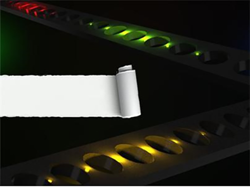
Figure 1. Illustration of photonic crystal lasers on paper substrates
Figure 2. Photonic crystal resonator laser and refractive index sensor operating on paper substrates
2017.03.01 View 7822
Semiconductor Photonic Nanocavities on Paper Substrates
Professor Yong-Hoon Cho of the Department of Physics and his team at KAIST have developed a semiconductor photonic nanocavity laser that can operate on a paper substrate.
The researchers hope that this novel method, which involves transferring nano-sized photonic crystal particles onto a paper substrate with high absorptiveness, will enable the diagnoses of various diseases by using high-tech semiconductor sensors at low cost.
The results of this research were published in the November 17th, 2016, issue of Advanced Materials.
Photonic crystals, which utilize light as a medium to provide high bandwidths, can transfer large amounts of information. Compared with their electronic counterparts, photonic crystals also consume less energy to operate.
Normally, semiconductor photonic particles require substrates, which play only a passive role in the assembly and endurance of individual, functional photonic components. These substrates, however, are bulky and environmentally hazardous as they are made up of non-biodegradable materials.
The research team overcame these two shortcomings by replacing a semiconductor substrate with standard paper. The substrate’s mass was reduced considerably, and because paper is made from trees, it degrades. Paper can be easily and cheaply acquired from our surroundings, which drastically reduces the unit cost of semiconductors.
In addition, paper possesses superior mechanical characteristics. It is flexible and can be repeatedly folded and unfolded without being torn. These are traits that have long been sought by researchers for existing flexible substrates.
The research team used a micro-sized stamp to detach photonic crystal nanobeam cavities selectively from their original substrate and transfer them onto a new paper substrate. Using this technique, the team removed nanophotonic crystals that had been patterned (using a process of selectively etching circuits onto a substrate) onto a semiconductor substrate with a high degree of integration, and realigned them as desired on a paper substrate.
The nanophotonic crystals that the team combined with paper in this research were 0.5 micrometers in width, 6 micrometers in length, and 0.3 micrometers in height—about one-hundredth of the width of a single hair (0.1 millimeter).
The team also transferred their photonic crystals onto paper with a fluid channel, which proved that it could be used as a refractive index sensor. As can be seen in current commercial pregnancy diagnosis kits, paper has high absorptiveness. Since photonic crystal particles have high sensitivity, they are highly suitable for applications such as sensors.
Professor Cho stated that “by using paper substrates, this technology can greatly contribute to the rising field of producing environmentally-friendly photonic particles” and “by combining inexpensive paper and high-performance photonic crystal sensors, we can obtain low prices as well as designing appropriate technologies with high performance.”
Dr. Sejeong Kim of the Department of Physics participated in this study as the first author, and Professor Kwanwoo Shin of Sogang University and Professor Yong-Hee Lee of KAIST also took part in this research. The research was supported by the National Research Foundation’s Mid-Career Researcher Program, and the Climate Change Research Hub of KAIST.
Figure 1. Illustration of photonic crystal lasers on paper substrates
Figure 2. Photonic crystal resonator laser and refractive index sensor operating on paper substrates
2017.03.01 View 7822 -
 A KAIST Alumnus Receives the Marie Sklodowska-Curie Individual Fellowships
Dr. Je-Kyung Ryu, a graduate of the Physics Department at KAIST in 2014, received the 2017 Marie Sklodowska-Curie Individual Fellowship.
Established in 1996, the Marie Sklodowska-Curie Individual Fellowships support young scientists in or outside Europe to help them grow as independent researchers. The recipients are recognized to have the highest potential to make a difference in science and technology and work on research and innovation.
Dr. Ryu is currently working as a postdoctoral researcher at the Cees Dekker Lab in the Department of Bionanoscience at the Kavli Institute of Nanoscience at Delft University of Technology (TU Delft), Netherlands.
He was among six international researchers at TU Delft who were awarded this research grant.
The grant of 177,000 euros will be offered for two years from March 2017 to February 2019 to cover his salary and research expenses.
For a news article published by TU Delft on the award, please click below:
QN and BN Successfully Attract Young Scientific Talent
February 1, 2017
2017.02.22 View 7101
A KAIST Alumnus Receives the Marie Sklodowska-Curie Individual Fellowships
Dr. Je-Kyung Ryu, a graduate of the Physics Department at KAIST in 2014, received the 2017 Marie Sklodowska-Curie Individual Fellowship.
Established in 1996, the Marie Sklodowska-Curie Individual Fellowships support young scientists in or outside Europe to help them grow as independent researchers. The recipients are recognized to have the highest potential to make a difference in science and technology and work on research and innovation.
Dr. Ryu is currently working as a postdoctoral researcher at the Cees Dekker Lab in the Department of Bionanoscience at the Kavli Institute of Nanoscience at Delft University of Technology (TU Delft), Netherlands.
He was among six international researchers at TU Delft who were awarded this research grant.
The grant of 177,000 euros will be offered for two years from March 2017 to February 2019 to cover his salary and research expenses.
For a news article published by TU Delft on the award, please click below:
QN and BN Successfully Attract Young Scientific Talent
February 1, 2017
2017.02.22 View 7101 -
 Dr. Sung-Chul Shin Selected 16th President of KAIST
(President Sung-Chul Shin)
The KAIST Board of Trustees elected Professor Sung-Chul Shin of the Department of Physics the 16th president of KAIST on February 21. Professor Shin succeeds President Sung-Mo Kang whose four-year term will end on February 23. He is the first KAIST alumnus to serve as its president.
The Board of Trustees announced, “We believe that Professor Shin’s scientific achievement, outstanding leadership, and clear vision will serve KAIST faculty, students, and staff very well. He will be the best person to help KAIST leap forward in the four years ahead.”
The newly-elected president said, “I am humbled and honored to have been elected to lead such a prestigious institute of Korea. Aiming to be one of the top ten global universities, KAIST will continue to innovate its systems.” Previously, Dr. Shin led the Daegu Gyeongbuk Institute of Science and Technology (DGIST) for six years as president since 2011.
Professor Shin joined the KAIST faculty in 1989. He graduated from Seoul National University and then earned his MS degree in condensed matter physics at KAIST in 1977. After earning his Ph.D. in material physics at Northwestern University in 1984, he worked at Eastman Kodak Research Labs as a senior research scientist for five years.
Before heading to DGIST, President Shin held key administrative positions at KAIST from the early 1990s including dean of planning, dean of the international office, and vice-dean of student affairs. During President Robert Laughlin’s tenure, the first foreign president at KAIST, he served as vice-president for two years from 2004. He also served on the Presidential Advisory Council on Science and Technology of the Korean government as vice chairperson from 2015 to 2016.
A renowned scholar in the field of nanoscience, President Shin’s research focuses on the artificial synthesis and characterization of nonmagnetic materials, magnetic anisotropy, and magneto-optical phenomena. He leads the Laboratory for Nanospinics of Spintronic Materials at KAIST and has published in 290 journals while holding 37 patents.
A fellow in the American Physical Society (APS) since 2008, he was the president of the Korean Physical Society from 2011 to 2012. He has been on the editorial board of J. Magnetism and Magnetic Materials from 2009 and was the first Korean recipient of the Asian Union of Magnetics Societies (AUMS) Award, which recognizes outstanding scientists in the field of magnetics.
President Shin envisions making KAIST’s research and education more competitive through continuing innovation. His innovation efforts will extend to the five key areas of education, research, technology commercialization, globalization, and future planning.
Among his priorities, he emphasizes multidisciplinary studies and leadership training for students. He plans to focus on undeclared major courses for undergraduates to help them expand their experience and exposure to diverse disciplines. This approach will help create well-rounded engineers, scientists, and entrepreneurs by enabling them to develop skills while leveraging a strong connection to the arts, humanities, and social sciences.
To better respond to Industry 4.0, which calls for convergence studies and collaborative work, he proposed establishing a ‘Convergence Innovation System’ by strategically selecting 10 flagship convergence research groups. In order to accelerate the technology commercialization and ecosystem of start-ups, he will strengthen entrepreneurship education, making it a prerequisite requirement for students. President Shin said he will spare no effort to incubate and spin-off ventures in which KAIST technology is being transferred. For globalization efforts, he plans to increase the ratio of foreign faculty from 9 percent to 15 percent, while doubling the current foreign student enrollment ratio of 5 percent. For future strategic innovation, he will implement a long-term innovation strategic plan dubbed ‘Vision 2031.’
2017.02.22 View 12068
Dr. Sung-Chul Shin Selected 16th President of KAIST
(President Sung-Chul Shin)
The KAIST Board of Trustees elected Professor Sung-Chul Shin of the Department of Physics the 16th president of KAIST on February 21. Professor Shin succeeds President Sung-Mo Kang whose four-year term will end on February 23. He is the first KAIST alumnus to serve as its president.
The Board of Trustees announced, “We believe that Professor Shin’s scientific achievement, outstanding leadership, and clear vision will serve KAIST faculty, students, and staff very well. He will be the best person to help KAIST leap forward in the four years ahead.”
The newly-elected president said, “I am humbled and honored to have been elected to lead such a prestigious institute of Korea. Aiming to be one of the top ten global universities, KAIST will continue to innovate its systems.” Previously, Dr. Shin led the Daegu Gyeongbuk Institute of Science and Technology (DGIST) for six years as president since 2011.
Professor Shin joined the KAIST faculty in 1989. He graduated from Seoul National University and then earned his MS degree in condensed matter physics at KAIST in 1977. After earning his Ph.D. in material physics at Northwestern University in 1984, he worked at Eastman Kodak Research Labs as a senior research scientist for five years.
Before heading to DGIST, President Shin held key administrative positions at KAIST from the early 1990s including dean of planning, dean of the international office, and vice-dean of student affairs. During President Robert Laughlin’s tenure, the first foreign president at KAIST, he served as vice-president for two years from 2004. He also served on the Presidential Advisory Council on Science and Technology of the Korean government as vice chairperson from 2015 to 2016.
A renowned scholar in the field of nanoscience, President Shin’s research focuses on the artificial synthesis and characterization of nonmagnetic materials, magnetic anisotropy, and magneto-optical phenomena. He leads the Laboratory for Nanospinics of Spintronic Materials at KAIST and has published in 290 journals while holding 37 patents.
A fellow in the American Physical Society (APS) since 2008, he was the president of the Korean Physical Society from 2011 to 2012. He has been on the editorial board of J. Magnetism and Magnetic Materials from 2009 and was the first Korean recipient of the Asian Union of Magnetics Societies (AUMS) Award, which recognizes outstanding scientists in the field of magnetics.
President Shin envisions making KAIST’s research and education more competitive through continuing innovation. His innovation efforts will extend to the five key areas of education, research, technology commercialization, globalization, and future planning.
Among his priorities, he emphasizes multidisciplinary studies and leadership training for students. He plans to focus on undeclared major courses for undergraduates to help them expand their experience and exposure to diverse disciplines. This approach will help create well-rounded engineers, scientists, and entrepreneurs by enabling them to develop skills while leveraging a strong connection to the arts, humanities, and social sciences.
To better respond to Industry 4.0, which calls for convergence studies and collaborative work, he proposed establishing a ‘Convergence Innovation System’ by strategically selecting 10 flagship convergence research groups. In order to accelerate the technology commercialization and ecosystem of start-ups, he will strengthen entrepreneurship education, making it a prerequisite requirement for students. President Shin said he will spare no effort to incubate and spin-off ventures in which KAIST technology is being transferred. For globalization efforts, he plans to increase the ratio of foreign faculty from 9 percent to 15 percent, while doubling the current foreign student enrollment ratio of 5 percent. For future strategic innovation, he will implement a long-term innovation strategic plan dubbed ‘Vision 2031.’
2017.02.22 View 12068 -
 The 2016 Research Highlights
KAIST has selected the ten most outstanding projects of 2016 conducted by its faculty and researchers. This selection embodies the KAIST research portfolios that translate their discoveries into meaningful and measurable impact toward a better world. All of them demonstrate exceptional creativity, which open new research paths for each field in its novelty, innovation, and impact.
The following list has been reviewed by a committee of faculty peers headed by Associate Vice President for Research. Following are the 2016 KAIST research highlights:
□ Commercialization of 3D Holographic Microscopy
By YongKeun Park of the Department of Physics
Professor YongKeun Park and his colleagues develop a powerful technique to measure 3D images of live cells without labeling agents. This technique, called 3D holographic microscopy or holotomography, will open a new avenue for the study of cell biology and its applications in medical diagnosis. This research also led to the founding of a start-up company Tomocube Inc. and the successful commercialization of the technique.
Professor Park and his research team developed a solution based on digital holography technology used to visualize 3D refractive index tomograms of live cells without staining. This allowed the real-time observation of biological cells in 2D, 3D, and 4D without the use of labeling agents. Conventional techniques for 3D cell imaging requires the use of labeling agents such as fluorescence dyes and proteins, which prevent from investigating the physiology of intact untreated cells. In particular, label-free imaging capability becomes more important in several emerging fields such as stem cell research and immunotherapy.
The team employs the concept of 3D digital holography to achieve the optical measurements of 3D refractive index tomograms of live cells and tissues. Also, a digital micromirror device (DMD), which has been used for DLPTM projectors, was utilized to steer a laser beam for 3D measurements.
Tomocube, founded from seed money funded by the EndRun Project of the Institute for Startup KAIST, succeeded in the commercialization of the 3D holographic microscopy and established an international distribution network in more than ten countries. It now has started exporting the product to several countries. The microscopes are being used in several leading research institutes including MIT, German Cancer Center, Pittsburg Medical Center, and Seoul National University Hospital Selected as one of the top ten mechanical technologies of 2016 by the Korean Society of Mechanical Engineers, the team raised four billion KRW investment from industry leaders including Soft Bank Venture Korea, Hanmi Pharmaceutical, and InterVest investment.
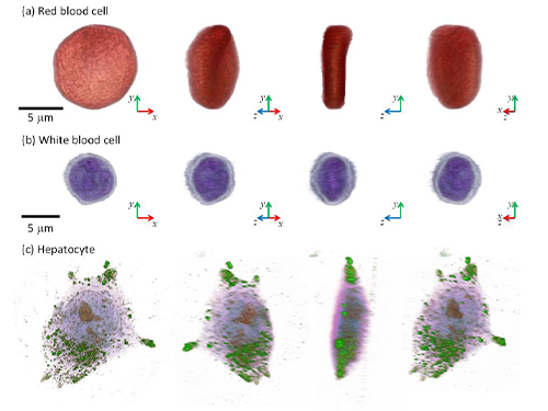
(Figure: Images of cells measured by 3D microscopy)
□ Designer Proteins with Chemical Modifications
By Hee-Sung Park and Hee Yoon Lee of the Department of Chemistry
Professor Hee-Sung Park developed a new strategy for installing authentic post-translational modifications (PTM) into recombinant proteins. Most essential biological processes are controlled by PTM, which plays a critical role in metabolic changes. However, abnormal protein modification aroused by environmental or genetic factors induce diverse diseases such as neurodegenerative diseases, cancer, and many other chronic diseases.
Professor Park has conceived a novel chemical biology route to achieve authentic and selective chemical modifications in proteins.He first used the established O-phosphoserine (Sep) orthogonal translational system to create a Sep-containing protein. The Sep residue is then dephosphorylated to dehydroalanine (Dha). Finally, Zn-Cu is conjugated to Dha of alkyl iodides, which enables it to form chemo-selective carbon-carbon bonds.
This approach offers a powerful tool to engineer designer proteins with diverse chemical modifications, providing a novel platform for investigating numerous diseases and drug development including for cancer and Alzheimer's. Furthermore, this research will allow mass production of abnormally modified proteins that could induce diseases, opening up new prospects in disease treatment research. It will help to enable investigation and discovery of new drug inhibitors that directly target abnormally modified proteins.
(Figure: Application of Customized Protein Modification Technology)
□ Lanthanum-Catalyzed Synthesis of Microporous 3D Graphene-Like Carbons in a Zeolite Template
By Ryong Ryoo, of the Department of Chemistry
Professor Ryong Ryoo’s team presented a scaled-up carbon synthesis viable for practical applications such as Li-ion batteries and catalyst supports.
Zeolite-templated carbon has an extremely large surface area and a regular microporous structure. As a result, it was expected to show excellent performance in various applications, such as for electrode materials or catalyst supports. However, until recently difficulties in synthesis have hindered research on application and properties of zeolited-templated carbon compared to other porous carbon materials.
Professor Ryoo’s team demonstrated that lanthanum ions embedded in zeolite pores lowered the temperature for carbonization of ethylene or acetylene. In this contribution, a graphene-like carbon structure was selectively formed inside zeolite template without the non-selective carbon deposition. Single crystal X-ray diffraction data revealed that carbon formed along the micropore surface. After removal of zeolite template, the carbon framework showed high electrical conductivity.
His synthesis method not only allowed selectivity in ethylene carbonization inside zeolite pore but permitted the diffusion of carbon material even when a large amount of zeolites was synthesized at once, allowing mass production of carbon. Thus, this method is expected to accelerate research on the application and properties of zeolite-templated carbon.
(Figure: Electron density distribution of zeolite that underwent selective pore carbonization. The structure of carbon determined by electron density distributions of carbon atoms, shown in yellow and red, within the framework of zeolite, shown in blue, can be observed.)
□ Complete Prevention of Blood Loss by Self-Sealing Hemostatic Needles
By Haeshin Lee of the Department of Chemistry
Professor Haeshin Lee’s team invented a hemostatic hypodermic needle, which prevented bleeding of punctured tissue during and after injections.
Bleeding unavoidably accompanies injections when a conventional needle penetrates tissue. Though the scale of bleeding from controlled injections does not cause harm to healthy individuals, uncontrolled bleeding may bring serious complications for those who suffer from hemophilia, coagulopathy, or who have been exposed to infectious diseases.
Professor Lee’s hemostatic hypodermic needle is coated with partially cross-linked catechol-functionalized chitosan that undergoes a solid-to-gel phase transition in situ to seal-seal punctured tissues. The team reported a complete prevention of blood loss following intravenous and intramuscular injections in animal models. They observed a 100% survival rate in hemophiliac mice following a syringe injection into a jugular vein.
The self-sealing hemostatic needles may help to prevent complications associated with bleeding in clinical settings such as for diabetic patients who experience delayed hemostasis and in the procedure of biopsy thereby preventing profuse bleeding.
□ An Immunological Mechanism for the Contribution of Commensal Microbiota Against Herpes Simplex Virus Infection in Genital Mucosa
By Heung Kyu Lee of the Graduate School of Medical Science and Engineering
Professor Heung Kyu Lee identified an immunological mechanism of commensal microbiota against herpes virus infections. The protective mechanisms of commensal bacteria against viral infections was limited to how immune inductive signals are provided by commensal bacteria for enhancing innate and adaptive immunity. Until Professor Lee’s research discovery, whether, or how, commensal bacteria might influence the effector arm of immune responses such as effector T cells to eliminate infected virus remained unknown.
Professor Lee’s team demonstrated that dysbiosis within the vaginal microbiota resulted in severe impairment of antiviral protection against HSV-2 infection. IL-33 released into the vaginal tract after antibiotic treatment blocked the ability of effector T cells to migrate into vaginal tissues and secrete the antiviral cytokine, IFN-γ. Thus, the findings suggested a previously unstudied role of commensal bacteria in the effector phase of the antiviral immune response against genital herpes. These findings provided insight into the mechanisms by which the secretion of proteases from opportunistic pathogens in susceptibility to various sexually transmitted pathogens might induce type 2 immunity within the female genital tract.
Promoting awareness of overuse of antibiotics, the research is expected to contribute to the development of viral vaccines with enhanced defense capacity by regulating commensal bacteria to promote health.
□ Development of a Pulse-Echo Laser Ultrasonic Propagation Imaging System
By Jung-Ryul Lee of the Department of Aerospace Engineering
Professor Jung-Ryul Lee’s team for the first time developed a mobile laser ultrasonic propagation imaging system that is capable of 2500-point inspection per second and visualization of pulse-echo ultrasonic wave through the thickness of a solid medium. This novel ultrasonic propagation visualization system has been successfully prototyped for the application of in-situ and in-process nondestructive evaluation of aerospace structures.
The real world proof-of-concept was achieved by testing the new system in the inspection of a space launcher fuselage (KSLV-II), control surfaces of military transport (CN-235), and the brake disk of F-16, guided weapon fuselage. In addition, the system has passed F-16 standard specimen test done by Korea Air Force and got a US patent. The prototype which was developed over a period of two years has been successfully delivered to Korea Air Force last December.
Furthermore, Boeing has expressed interest in prototype development project and KAIST OESL has been selected as the Boeing-KAIST technical contact lab and received a two-year grant from Boeing. The second prototype is under construction for Boeing and the third prototype will be delivered to an optional research institute and used as a standard inspection instrument.
□ Birefractive Stereo Imaging for Single-Shot Depth Acquisition
By Min H. Kim of the School of Computing
Professor Min H. Kim’s team proposed a novel 3D imaging method that allows the capture of not only color pictures but also corresponding depth images while traditional cameras capture just color pictures.
Depending on the polarization state of light, the incident light on a birefringent material such as calcite can be refracted into two different angles. This physical phenomenon is called double refraction. Whereas traditional stereo imaging requires at least two stereo cameras, 3D imaging method can capture depth from a single picture of double refraction. This proposed 3D imaging technique can be applied to many graphics and computer vision applications such as AR/VR applications that require color and depth information simultaneously.
This technology, which could measure depth images, is currently needed for various industrial applications. The suggested method in this research to measure depth information from one photo using double refraction media accurately can be used in areas where system size and cost are important, such as mobile cameras, VR/ARs, driverless cars, and 3D microscopes.
(Figure: Measuring high-resolution depth of single image via bi-refringent medium)
□Development of Environment Friendly Geotechnical Construction Material Using Biopolymer
By Gye-Chun Cho of the Department of Civil and Environmental Engineering
Professor Gye-Chun Cho has succeeded in making a 100% bio-based KABS (KAIST Bio-Soil) binder using biopolymer, an eco-friendly geotechnical construction material. A biopolymer is an organic polymer produced in the course of microbial activities and thus is an eco-friendly material manufactured without generating carbon dioxide. Biopolymers have been used in food, agriculture, cosmetics, and medicine as hardener and gelling agents, but have never been applied in construction. His team verified the microscopic interaction, feasibility, and strengthening mechanism of microbial biopolymers for soils for the first time in the world, suggesting that biopolymers be an eco-friendly soil binder.
In addition to soil binders, biopolymers can also be applied to various fields of ground construction (e.g., ground improvement, grouting, erosion control, vegetation, anti-desertification, etc.). The team expects more biopolymer applications in construction since increasing demands for replacing cement-based or chemical ground materials have surged.
With KABS binder, the team has performed several field tests along with industrial technology transfer underway. In collaboration with the Korea Expressway Corporation and LH Corporation, Professor Cho’s team is working on additional commercial applications.
(Figure: Strength enhancement effect of soil grain processed by biopolymer )
□ Protein Delivery Via Engineered Exosomes
By Chulhee Choi of the Department of Bio and Brain Engineering
Professor Chulhee Choi’s team unveiled a new tool for intracellular delivery of target proteins, named “exosomes for protein loading via optically reversible protein-protein interactions” or “EXPLORs”.
Nanoparticle-mediated delivery of functional macromolecules is a promising method for treating a variety of human diseases. Among nanoparticles, cell-derived exosomes have recently gained attention as a new therapeutic strategy for the in vivo delivery of nucleotides and chemical drugs.
By integrating a reversible protein-protein interaction module controlled by blue light with the endogenous process of exosome biogenesis, the team successfully loaded cargo proteins into newly generated exosomes. Treatment with protein-loaded EXPLORs is shown to significantly increase intracellular levels of cargo proteins and their function in recipient cells in vitro and in vivo.
These results clearly indicate the potential of EXPLORs as a mechanism for the efficient intracellular transfer of protein-based therapeutics into recipient cells and tissues. This technology has been transferred to KAIST bio-venture Cellex Life Science, Incorporated for commercialization.
□ Hot Electron Detection under Catalytic Reactions
By Jeong Young Park of the Graduate School of EEWS
Professor Jeong Young Park’s team developed a novel catalytic nanodiode consisting of a thin metal catalyst deposited onto a semiconductor support. The team succeeded in observing in real-time hot electrons created in the course of catalytic reaction occurring at atmospheric pressure or at liquid-solid interfaces.
Use of a noble catalytic nanodiode is a new measurement system that detects hot electrons produced on catalyst surface through atmospheric pressure and liquid chemical reaction in real time that allows direct identification of the catalytic activity of catalytic reactions. In particular, the system allows macro-observation of hot-electron movements that change with the type of nano-catalyst without high-priced equipment in atmospheric pressure and liquidation, and thus is not limited to experimental conditions such as in ultrahigh vacuums.
Therefore, it could be applied in the future to analyze complex chemical reaction mechanisms of catalysts used in high temperature and various pressure conditions, and to develop high efficiency next-generation catalyst materials. This finding may lead not only to the fundamental understanding in the mechanism of the catalytic reactions but also to the development of next-generation catalysts with enhanced catalytic performance.
(Figure: Schematic diagrams of nano-catalyst hot electron element and graphene hot electron detector)
2017.02.20 View 13899
The 2016 Research Highlights
KAIST has selected the ten most outstanding projects of 2016 conducted by its faculty and researchers. This selection embodies the KAIST research portfolios that translate their discoveries into meaningful and measurable impact toward a better world. All of them demonstrate exceptional creativity, which open new research paths for each field in its novelty, innovation, and impact.
The following list has been reviewed by a committee of faculty peers headed by Associate Vice President for Research. Following are the 2016 KAIST research highlights:
□ Commercialization of 3D Holographic Microscopy
By YongKeun Park of the Department of Physics
Professor YongKeun Park and his colleagues develop a powerful technique to measure 3D images of live cells without labeling agents. This technique, called 3D holographic microscopy or holotomography, will open a new avenue for the study of cell biology and its applications in medical diagnosis. This research also led to the founding of a start-up company Tomocube Inc. and the successful commercialization of the technique.
Professor Park and his research team developed a solution based on digital holography technology used to visualize 3D refractive index tomograms of live cells without staining. This allowed the real-time observation of biological cells in 2D, 3D, and 4D without the use of labeling agents. Conventional techniques for 3D cell imaging requires the use of labeling agents such as fluorescence dyes and proteins, which prevent from investigating the physiology of intact untreated cells. In particular, label-free imaging capability becomes more important in several emerging fields such as stem cell research and immunotherapy.
The team employs the concept of 3D digital holography to achieve the optical measurements of 3D refractive index tomograms of live cells and tissues. Also, a digital micromirror device (DMD), which has been used for DLPTM projectors, was utilized to steer a laser beam for 3D measurements.
Tomocube, founded from seed money funded by the EndRun Project of the Institute for Startup KAIST, succeeded in the commercialization of the 3D holographic microscopy and established an international distribution network in more than ten countries. It now has started exporting the product to several countries. The microscopes are being used in several leading research institutes including MIT, German Cancer Center, Pittsburg Medical Center, and Seoul National University Hospital Selected as one of the top ten mechanical technologies of 2016 by the Korean Society of Mechanical Engineers, the team raised four billion KRW investment from industry leaders including Soft Bank Venture Korea, Hanmi Pharmaceutical, and InterVest investment.
(Figure: Images of cells measured by 3D microscopy)
□ Designer Proteins with Chemical Modifications
By Hee-Sung Park and Hee Yoon Lee of the Department of Chemistry
Professor Hee-Sung Park developed a new strategy for installing authentic post-translational modifications (PTM) into recombinant proteins. Most essential biological processes are controlled by PTM, which plays a critical role in metabolic changes. However, abnormal protein modification aroused by environmental or genetic factors induce diverse diseases such as neurodegenerative diseases, cancer, and many other chronic diseases.
Professor Park has conceived a novel chemical biology route to achieve authentic and selective chemical modifications in proteins.He first used the established O-phosphoserine (Sep) orthogonal translational system to create a Sep-containing protein. The Sep residue is then dephosphorylated to dehydroalanine (Dha). Finally, Zn-Cu is conjugated to Dha of alkyl iodides, which enables it to form chemo-selective carbon-carbon bonds.
This approach offers a powerful tool to engineer designer proteins with diverse chemical modifications, providing a novel platform for investigating numerous diseases and drug development including for cancer and Alzheimer's. Furthermore, this research will allow mass production of abnormally modified proteins that could induce diseases, opening up new prospects in disease treatment research. It will help to enable investigation and discovery of new drug inhibitors that directly target abnormally modified proteins.
(Figure: Application of Customized Protein Modification Technology)
□ Lanthanum-Catalyzed Synthesis of Microporous 3D Graphene-Like Carbons in a Zeolite Template
By Ryong Ryoo, of the Department of Chemistry
Professor Ryong Ryoo’s team presented a scaled-up carbon synthesis viable for practical applications such as Li-ion batteries and catalyst supports.
Zeolite-templated carbon has an extremely large surface area and a regular microporous structure. As a result, it was expected to show excellent performance in various applications, such as for electrode materials or catalyst supports. However, until recently difficulties in synthesis have hindered research on application and properties of zeolited-templated carbon compared to other porous carbon materials.
Professor Ryoo’s team demonstrated that lanthanum ions embedded in zeolite pores lowered the temperature for carbonization of ethylene or acetylene. In this contribution, a graphene-like carbon structure was selectively formed inside zeolite template without the non-selective carbon deposition. Single crystal X-ray diffraction data revealed that carbon formed along the micropore surface. After removal of zeolite template, the carbon framework showed high electrical conductivity.
His synthesis method not only allowed selectivity in ethylene carbonization inside zeolite pore but permitted the diffusion of carbon material even when a large amount of zeolites was synthesized at once, allowing mass production of carbon. Thus, this method is expected to accelerate research on the application and properties of zeolite-templated carbon.
(Figure: Electron density distribution of zeolite that underwent selective pore carbonization. The structure of carbon determined by electron density distributions of carbon atoms, shown in yellow and red, within the framework of zeolite, shown in blue, can be observed.)
□ Complete Prevention of Blood Loss by Self-Sealing Hemostatic Needles
By Haeshin Lee of the Department of Chemistry
Professor Haeshin Lee’s team invented a hemostatic hypodermic needle, which prevented bleeding of punctured tissue during and after injections.
Bleeding unavoidably accompanies injections when a conventional needle penetrates tissue. Though the scale of bleeding from controlled injections does not cause harm to healthy individuals, uncontrolled bleeding may bring serious complications for those who suffer from hemophilia, coagulopathy, or who have been exposed to infectious diseases.
Professor Lee’s hemostatic hypodermic needle is coated with partially cross-linked catechol-functionalized chitosan that undergoes a solid-to-gel phase transition in situ to seal-seal punctured tissues. The team reported a complete prevention of blood loss following intravenous and intramuscular injections in animal models. They observed a 100% survival rate in hemophiliac mice following a syringe injection into a jugular vein.
The self-sealing hemostatic needles may help to prevent complications associated with bleeding in clinical settings such as for diabetic patients who experience delayed hemostasis and in the procedure of biopsy thereby preventing profuse bleeding.
□ An Immunological Mechanism for the Contribution of Commensal Microbiota Against Herpes Simplex Virus Infection in Genital Mucosa
By Heung Kyu Lee of the Graduate School of Medical Science and Engineering
Professor Heung Kyu Lee identified an immunological mechanism of commensal microbiota against herpes virus infections. The protective mechanisms of commensal bacteria against viral infections was limited to how immune inductive signals are provided by commensal bacteria for enhancing innate and adaptive immunity. Until Professor Lee’s research discovery, whether, or how, commensal bacteria might influence the effector arm of immune responses such as effector T cells to eliminate infected virus remained unknown.
Professor Lee’s team demonstrated that dysbiosis within the vaginal microbiota resulted in severe impairment of antiviral protection against HSV-2 infection. IL-33 released into the vaginal tract after antibiotic treatment blocked the ability of effector T cells to migrate into vaginal tissues and secrete the antiviral cytokine, IFN-γ. Thus, the findings suggested a previously unstudied role of commensal bacteria in the effector phase of the antiviral immune response against genital herpes. These findings provided insight into the mechanisms by which the secretion of proteases from opportunistic pathogens in susceptibility to various sexually transmitted pathogens might induce type 2 immunity within the female genital tract.
Promoting awareness of overuse of antibiotics, the research is expected to contribute to the development of viral vaccines with enhanced defense capacity by regulating commensal bacteria to promote health.
□ Development of a Pulse-Echo Laser Ultrasonic Propagation Imaging System
By Jung-Ryul Lee of the Department of Aerospace Engineering
Professor Jung-Ryul Lee’s team for the first time developed a mobile laser ultrasonic propagation imaging system that is capable of 2500-point inspection per second and visualization of pulse-echo ultrasonic wave through the thickness of a solid medium. This novel ultrasonic propagation visualization system has been successfully prototyped for the application of in-situ and in-process nondestructive evaluation of aerospace structures.
The real world proof-of-concept was achieved by testing the new system in the inspection of a space launcher fuselage (KSLV-II), control surfaces of military transport (CN-235), and the brake disk of F-16, guided weapon fuselage. In addition, the system has passed F-16 standard specimen test done by Korea Air Force and got a US patent. The prototype which was developed over a period of two years has been successfully delivered to Korea Air Force last December.
Furthermore, Boeing has expressed interest in prototype development project and KAIST OESL has been selected as the Boeing-KAIST technical contact lab and received a two-year grant from Boeing. The second prototype is under construction for Boeing and the third prototype will be delivered to an optional research institute and used as a standard inspection instrument.
□ Birefractive Stereo Imaging for Single-Shot Depth Acquisition
By Min H. Kim of the School of Computing
Professor Min H. Kim’s team proposed a novel 3D imaging method that allows the capture of not only color pictures but also corresponding depth images while traditional cameras capture just color pictures.
Depending on the polarization state of light, the incident light on a birefringent material such as calcite can be refracted into two different angles. This physical phenomenon is called double refraction. Whereas traditional stereo imaging requires at least two stereo cameras, 3D imaging method can capture depth from a single picture of double refraction. This proposed 3D imaging technique can be applied to many graphics and computer vision applications such as AR/VR applications that require color and depth information simultaneously.
This technology, which could measure depth images, is currently needed for various industrial applications. The suggested method in this research to measure depth information from one photo using double refraction media accurately can be used in areas where system size and cost are important, such as mobile cameras, VR/ARs, driverless cars, and 3D microscopes.
(Figure: Measuring high-resolution depth of single image via bi-refringent medium)
□Development of Environment Friendly Geotechnical Construction Material Using Biopolymer
By Gye-Chun Cho of the Department of Civil and Environmental Engineering
Professor Gye-Chun Cho has succeeded in making a 100% bio-based KABS (KAIST Bio-Soil) binder using biopolymer, an eco-friendly geotechnical construction material. A biopolymer is an organic polymer produced in the course of microbial activities and thus is an eco-friendly material manufactured without generating carbon dioxide. Biopolymers have been used in food, agriculture, cosmetics, and medicine as hardener and gelling agents, but have never been applied in construction. His team verified the microscopic interaction, feasibility, and strengthening mechanism of microbial biopolymers for soils for the first time in the world, suggesting that biopolymers be an eco-friendly soil binder.
In addition to soil binders, biopolymers can also be applied to various fields of ground construction (e.g., ground improvement, grouting, erosion control, vegetation, anti-desertification, etc.). The team expects more biopolymer applications in construction since increasing demands for replacing cement-based or chemical ground materials have surged.
With KABS binder, the team has performed several field tests along with industrial technology transfer underway. In collaboration with the Korea Expressway Corporation and LH Corporation, Professor Cho’s team is working on additional commercial applications.
(Figure: Strength enhancement effect of soil grain processed by biopolymer )
□ Protein Delivery Via Engineered Exosomes
By Chulhee Choi of the Department of Bio and Brain Engineering
Professor Chulhee Choi’s team unveiled a new tool for intracellular delivery of target proteins, named “exosomes for protein loading via optically reversible protein-protein interactions” or “EXPLORs”.
Nanoparticle-mediated delivery of functional macromolecules is a promising method for treating a variety of human diseases. Among nanoparticles, cell-derived exosomes have recently gained attention as a new therapeutic strategy for the in vivo delivery of nucleotides and chemical drugs.
By integrating a reversible protein-protein interaction module controlled by blue light with the endogenous process of exosome biogenesis, the team successfully loaded cargo proteins into newly generated exosomes. Treatment with protein-loaded EXPLORs is shown to significantly increase intracellular levels of cargo proteins and their function in recipient cells in vitro and in vivo.
These results clearly indicate the potential of EXPLORs as a mechanism for the efficient intracellular transfer of protein-based therapeutics into recipient cells and tissues. This technology has been transferred to KAIST bio-venture Cellex Life Science, Incorporated for commercialization.
□ Hot Electron Detection under Catalytic Reactions
By Jeong Young Park of the Graduate School of EEWS
Professor Jeong Young Park’s team developed a novel catalytic nanodiode consisting of a thin metal catalyst deposited onto a semiconductor support. The team succeeded in observing in real-time hot electrons created in the course of catalytic reaction occurring at atmospheric pressure or at liquid-solid interfaces.
Use of a noble catalytic nanodiode is a new measurement system that detects hot electrons produced on catalyst surface through atmospheric pressure and liquid chemical reaction in real time that allows direct identification of the catalytic activity of catalytic reactions. In particular, the system allows macro-observation of hot-electron movements that change with the type of nano-catalyst without high-priced equipment in atmospheric pressure and liquidation, and thus is not limited to experimental conditions such as in ultrahigh vacuums.
Therefore, it could be applied in the future to analyze complex chemical reaction mechanisms of catalysts used in high temperature and various pressure conditions, and to develop high efficiency next-generation catalyst materials. This finding may lead not only to the fundamental understanding in the mechanism of the catalytic reactions but also to the development of next-generation catalysts with enhanced catalytic performance.
(Figure: Schematic diagrams of nano-catalyst hot electron element and graphene hot electron detector)
2017.02.20 View 13899 -
 A New Approach to 3D Holographic Displays Greatly Improves the Image Quality
With the addition of holographic diffusers or frosted glasses to wavefront modulators, KAIST researchers offer a simple and practical solution to significantly enhance the performance of 3D dynamic holographic displays by 2,600 times.
The potential applications of three-dimensional (3D) digital holograms are enormous. In addition to arts and entertainment, various fields including biomedical imaging, scientific visualization, engineering design, and displays could benefit from this technology. For example, creating full-sized organs for 3D analysis by doctors could be helpful, but it remained a challenge owing to the limitation of hologram-generation techniques.
A research team led by Professor YongKeun Park of the Physics Department at KAIST has come up with a solution and developed a 3D holographic display that performs more than 2,600 times better than existing 3D holographic displays. This study is expected to improve the limited size and viewing angle of 3D images, which were a major problem of the current holographic displays. The study was published online in Nature Photonics on January 23, 2017.
3D holograms, which often appear in science fiction films, are a familiar technology to the public, but holograms in movies are created with computer graphic effects. Methods for creating true 3D holograms are still being studied in the laboratory. For example, due to the difficulty of generating real 3D images, recent virtual reality (VR) and augmented reality (AR) devices project two different two-dimensional (2D) images onto a viewer to induce optical illusions.
To create a 3D hologram that can be viewed without special equipment such as 3D glasses, the wavefront of light must be controlled using wavefront modulators such as spatial light modulators (SLMs) and deformable mirrors (DMs). A wavefront modulator is an optical manipulation device that can control the direction of light propagation.
However, the biggest limitation to using these modulators as 3D displays is the number of pixels. The large number of pixels that are packed into high-resolution displays developed in recent years are suitable for a 2D image, and the amount of information contained in those pixels cannot produce a 3D image. For this reason, a 3D image that can be made with existing wavefront modulator technology is 1 cm in size with a narrow viewing angle of 3 degrees, which is far from practicable.
As an alternative, KAIST researchers used a DM and added two successive holographic diffusers to scatter light. By scattering light in many directions, this allows for a wider viewing angle and larger image, but results in volume speckle fields, which are caused by the interference of multiple scattered light. Random volume speckle fields cannot be used to display 3D images.
To fix the problem, the researchers employed a wavefront-shaping technique to control the fields. As a result, they succeeded in producing an enhanced 3D holographic image with a viewing angle of 35 degrees in a volume of 2 cm in length, width, and height. This yielded a performance that was about 2,600 times stronger than the original image definition generated when they used a DM without a diffuser.
Professor Park said, “Scattering light has previously been believed to interfere with the recognition of objects, but we have demonstrated that current 3D displays can be improved significantly with an increased viewing angle and image size by properly controlling the scattered light.”
Hyeonseung Yu, who is the lead author of this research article and a doctoral candidate in the Department of Physics, KAIST, noted that this technology signals a good start to develop a practical model for dynamic 3D hologram displays that can be enjoyed without the need for special eyeglasses. “This approach can also be applied to AR and VR technology to enhance the image resolution and viewing angles,” added Yu.
The research paper is entitled “Ultrahigh-definition Dynamic 3D Holographic Display by Active Control of Volume Speckle Fields.”
Figure 1. Concept of Scattering Display
The size and viewing angle of 3D images can be simultaneously increased when a scattering medium (diffuser) is introduced. By controlling the wavefront impinging on the scattering medium, the desired 3D hologram is generated.
Figure 2. Experimental Setup
The optical set-up consists of a deformable mirror and the scattering medium with two successive holographic diffusers. A high-numerical-aperture imaging unit mounted on a three-axis motorized translational system is utilized for wavefront optimization and imaging.
Figure 3. 3D Images Projected
This picture shows 3D images in a volume of 2 cm × 2 cm × 2 cm with a viewing angle of 35 degrees using one of the wavefront modulators, a digital micromirror device (DMD).
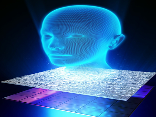
Figure 4. Artist’s Rendition of the Proposed Concept
A dynamic 3D hologram of a face is displayed.
2017.02.01 View 14027
A New Approach to 3D Holographic Displays Greatly Improves the Image Quality
With the addition of holographic diffusers or frosted glasses to wavefront modulators, KAIST researchers offer a simple and practical solution to significantly enhance the performance of 3D dynamic holographic displays by 2,600 times.
The potential applications of three-dimensional (3D) digital holograms are enormous. In addition to arts and entertainment, various fields including biomedical imaging, scientific visualization, engineering design, and displays could benefit from this technology. For example, creating full-sized organs for 3D analysis by doctors could be helpful, but it remained a challenge owing to the limitation of hologram-generation techniques.
A research team led by Professor YongKeun Park of the Physics Department at KAIST has come up with a solution and developed a 3D holographic display that performs more than 2,600 times better than existing 3D holographic displays. This study is expected to improve the limited size and viewing angle of 3D images, which were a major problem of the current holographic displays. The study was published online in Nature Photonics on January 23, 2017.
3D holograms, which often appear in science fiction films, are a familiar technology to the public, but holograms in movies are created with computer graphic effects. Methods for creating true 3D holograms are still being studied in the laboratory. For example, due to the difficulty of generating real 3D images, recent virtual reality (VR) and augmented reality (AR) devices project two different two-dimensional (2D) images onto a viewer to induce optical illusions.
To create a 3D hologram that can be viewed without special equipment such as 3D glasses, the wavefront of light must be controlled using wavefront modulators such as spatial light modulators (SLMs) and deformable mirrors (DMs). A wavefront modulator is an optical manipulation device that can control the direction of light propagation.
However, the biggest limitation to using these modulators as 3D displays is the number of pixels. The large number of pixels that are packed into high-resolution displays developed in recent years are suitable for a 2D image, and the amount of information contained in those pixels cannot produce a 3D image. For this reason, a 3D image that can be made with existing wavefront modulator technology is 1 cm in size with a narrow viewing angle of 3 degrees, which is far from practicable.
As an alternative, KAIST researchers used a DM and added two successive holographic diffusers to scatter light. By scattering light in many directions, this allows for a wider viewing angle and larger image, but results in volume speckle fields, which are caused by the interference of multiple scattered light. Random volume speckle fields cannot be used to display 3D images.
To fix the problem, the researchers employed a wavefront-shaping technique to control the fields. As a result, they succeeded in producing an enhanced 3D holographic image with a viewing angle of 35 degrees in a volume of 2 cm in length, width, and height. This yielded a performance that was about 2,600 times stronger than the original image definition generated when they used a DM without a diffuser.
Professor Park said, “Scattering light has previously been believed to interfere with the recognition of objects, but we have demonstrated that current 3D displays can be improved significantly with an increased viewing angle and image size by properly controlling the scattered light.”
Hyeonseung Yu, who is the lead author of this research article and a doctoral candidate in the Department of Physics, KAIST, noted that this technology signals a good start to develop a practical model for dynamic 3D hologram displays that can be enjoyed without the need for special eyeglasses. “This approach can also be applied to AR and VR technology to enhance the image resolution and viewing angles,” added Yu.
The research paper is entitled “Ultrahigh-definition Dynamic 3D Holographic Display by Active Control of Volume Speckle Fields.”
Figure 1. Concept of Scattering Display
The size and viewing angle of 3D images can be simultaneously increased when a scattering medium (diffuser) is introduced. By controlling the wavefront impinging on the scattering medium, the desired 3D hologram is generated.
Figure 2. Experimental Setup
The optical set-up consists of a deformable mirror and the scattering medium with two successive holographic diffusers. A high-numerical-aperture imaging unit mounted on a three-axis motorized translational system is utilized for wavefront optimization and imaging.
Figure 3. 3D Images Projected
This picture shows 3D images in a volume of 2 cm × 2 cm × 2 cm with a viewing angle of 35 degrees using one of the wavefront modulators, a digital micromirror device (DMD).
Figure 4. Artist’s Rendition of the Proposed Concept
A dynamic 3D hologram of a face is displayed.
2017.02.01 View 14027 -
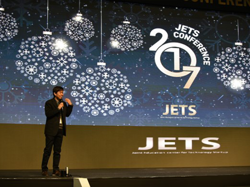 JETS Conference 2017
KAIST and four science and technology research universities in Korea co-hosted a technology start-up fair, the 2017 JETS (Job, Exhibition, Tech Forum, and Startup) Conference January 19 ~20 in the Ryu Geun-chul Sports Complex at KAIST.
Korea’s major science and technology research universities, Daegu Gyeongbuk Institute of Science and Technology (DGIST), Gwangju Institute of Science and Technology (GIST), Pohang University of Science and Technology (Postech), and Ulsan National Institute of Science and Technology (UNIST), held the event in a collaborative effort to educate, inspire, and connect young entrepreneurs, especially those who will launch technology start-ups.
The conference brought entrepreneurs and innovators together who seek ways of working with and supporting start-ups and for their sustainable growth. It also drew aspiring young students and researchers from universities and the government-funded research institutions who are in the process of commercializing their technology. Students from each university’s industry-academia cooperation program who incubated their technology and ideas were key contributors.
At the Tech Forum, entrepreneurship and technology consultation specialists including Joe Jasin, managing director at DNA Investment Partners in the US, the founder of Cyworld Dong-Hyung Lee, and Professor Hawoong Jeong, a complex bio-network specialist from the Department of Physics of KAIST lectured on the ecosystem of start-ups and its trends and development.
The Dean of University-Industry Cooperation at KAIST Joongmyeon Bae said, "We organized this event in collaboration with four major research universities to further encourage technology start-ups from young students and help their ideas and technology bear fruit. We will continue to strive to create an ecosystem of start-ups which works efficiently.”
(Above photo: Founder of the Cyworld, Dong-Hyung Lee gives a lecture at the Tech Forum. Below photo: Students visit exhibition booth of each participating institution.)
2017.01.20 View 12754
JETS Conference 2017
KAIST and four science and technology research universities in Korea co-hosted a technology start-up fair, the 2017 JETS (Job, Exhibition, Tech Forum, and Startup) Conference January 19 ~20 in the Ryu Geun-chul Sports Complex at KAIST.
Korea’s major science and technology research universities, Daegu Gyeongbuk Institute of Science and Technology (DGIST), Gwangju Institute of Science and Technology (GIST), Pohang University of Science and Technology (Postech), and Ulsan National Institute of Science and Technology (UNIST), held the event in a collaborative effort to educate, inspire, and connect young entrepreneurs, especially those who will launch technology start-ups.
The conference brought entrepreneurs and innovators together who seek ways of working with and supporting start-ups and for their sustainable growth. It also drew aspiring young students and researchers from universities and the government-funded research institutions who are in the process of commercializing their technology. Students from each university’s industry-academia cooperation program who incubated their technology and ideas were key contributors.
At the Tech Forum, entrepreneurship and technology consultation specialists including Joe Jasin, managing director at DNA Investment Partners in the US, the founder of Cyworld Dong-Hyung Lee, and Professor Hawoong Jeong, a complex bio-network specialist from the Department of Physics of KAIST lectured on the ecosystem of start-ups and its trends and development.
The Dean of University-Industry Cooperation at KAIST Joongmyeon Bae said, "We organized this event in collaboration with four major research universities to further encourage technology start-ups from young students and help their ideas and technology bear fruit. We will continue to strive to create an ecosystem of start-ups which works efficiently.”
(Above photo: Founder of the Cyworld, Dong-Hyung Lee gives a lecture at the Tech Forum. Below photo: Students visit exhibition booth of each participating institution.)
2017.01.20 View 12754 -
 Nobel Laureate Dr. John Michael Kosterlitz Speaks at KAIST
KAIST’s Department of Physics will invite one of three co-recipients of the Nobel Prize in Physics 2016, Professor John Michael Kosterlitz of Brown University, on January 9, 2017, to speak about the exotic states of matter, which is entitled “Topological Defects and Phase Transitions.”
Professor Kosterlitz shares the Nobel award with two other researchers, David Thouless and Duncan Haldane. He is considered one of the pioneers in the field of topological phases. In the early 1970s, along with Thouless, he demonstrated that superconductivity could occur at low temperatures and explained the mechanism behind, phase transition, that makes superconductivity disappear at higher temperatures.
Over the last decade, topological materials and their applications have been widely studied with the hope of using them in new generations of electronics and superconductors, or in future quantum computers. Details of the lecture follow below:
Distinguished Lecture Series by KAIST’s Physics Department
· Speaker: Professor John Michael Kosterlitz of the Physics Department,
Brown University
· Topic: “Topological Defects and Phase Transitions”
· Date: January 9, 2017, 4:00 PM
· Place: Lecture Hall (#1501), College of Natural Sciences (E6-2)
2017.01.06 View 7932
Nobel Laureate Dr. John Michael Kosterlitz Speaks at KAIST
KAIST’s Department of Physics will invite one of three co-recipients of the Nobel Prize in Physics 2016, Professor John Michael Kosterlitz of Brown University, on January 9, 2017, to speak about the exotic states of matter, which is entitled “Topological Defects and Phase Transitions.”
Professor Kosterlitz shares the Nobel award with two other researchers, David Thouless and Duncan Haldane. He is considered one of the pioneers in the field of topological phases. In the early 1970s, along with Thouless, he demonstrated that superconductivity could occur at low temperatures and explained the mechanism behind, phase transition, that makes superconductivity disappear at higher temperatures.
Over the last decade, topological materials and their applications have been widely studied with the hope of using them in new generations of electronics and superconductors, or in future quantum computers. Details of the lecture follow below:
Distinguished Lecture Series by KAIST’s Physics Department
· Speaker: Professor John Michael Kosterlitz of the Physics Department,
Brown University
· Topic: “Topological Defects and Phase Transitions”
· Date: January 9, 2017, 4:00 PM
· Place: Lecture Hall (#1501), College of Natural Sciences (E6-2)
2017.01.06 View 7932 -
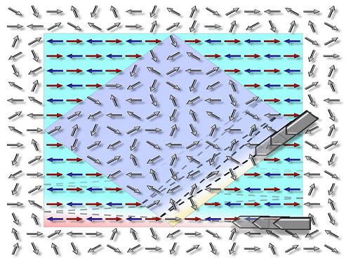 Technology to Allow Non-Magnetic Materials to Have Magnetic Properties by Professor Chan-Ho Yang
Professor Chan-Ho Yang and his research team from the Department of Physics at KAIST have developed a technology that allows non-magnetic materials to have magnetic properties or, in reverse, to remove magnetic properties from a magnet using an electric field.
Based on this research, it is expected that if magnetic-material-based data storage is developed, applications for high-speed massive data transfer will be possible.
The results of this research, with Ph.D. candidate Byung-Kwon Jang as the first author, were published online in Nature Physics on October 3.
Very small magnets exist inside of any materials. If the direction of the minuscule magnets is dis-aligned, pointing multiple directions, it is non-magnetic. If the direction is aligned in a certain direction, the material holds magnetic property just like any magnet we normally see.
Data storage capacity technology has rapidly advanced to the point where we can easily get a portable hard disk drive (HDD) with terabyte-level storage; however, the increase in storage is inevitably followed by slower data access speed for a storage device. Although HDDs are currently the most widely used data storage devices, their technical applications are limited due to their slow data access speed.
Other methods such as solid-state drives (SSDs), floating gates, and resistive switching have been developed as alternatives. Yet, they leave tracks every time data is written, and this can cause fatigue cumulative damage.
There have been many attempts to compose cells—the smallest data storage space on a storage device—with magnetic materials as that would enable faster data access speeds and remove fatigue cumulative damage. Generally, the techniques tried by researchers were to use induced magnetic fields through current flow. However, magnetic fields are very difficult to shield and can affect a large area. As a result, they alternate the magnetic property of adjacent cells. Because each cell cannot be adjusted one by one, it cannot also be arranged in a certain direction, and therefore, it is hard to change the magnetic state.
Professor Yang and his team adjusted the magnetic state by using magnetoelectric interaction to deal with this issue. Instead of using magnetic fields, magnetoelectric interaction is a method that uses an electric field to adjust the magnetic state. It has the advantage of smaller energy consumption as well.
Professor Yang's team demonstrated that cells facing random directions can be arranged in a certain direction by only inducing an electric field. In addition, the reverse was also proved to be feasible.
Until this research, most cases of previous findings were only feasible at extremely low temperatures or high temperatures, but the technology developed by the research team is practicable at room temperature by manipulating chemical pressure. It allows for a reversible magnetic state, and moreover, is non-volatile. Therefore, the results of this research are expected to provide the basis for developing next-generation information storage device.
Professor Yang said, “The changes in the electric magnetic state will be accompanied by entropy changes” and added, “Our research is expected to open new potential for future applications not only for magnetoelectric devices, but also for thermoelectric effect.”
This research has been worked on jointly with Dr. Si-Yong Choi from the Korea Institute of Materials Science, Prof. Yoon-Hee Jeong from the Pohang University of Science and Technology, Dr. Tae-Yeong Koo from the Pohang Accelerator Laboratory, Dr. Kyung-Tae Ko from the Max Planck Institute for Chemical Physics of Solids, Dr. Jun-Sik Lee and Dr. Hendrik Ohldag from the SLAC National Accelerator Laboratory of the United States, and Prof. Jan Seidel from the University of New South Wales of Australia.
The research was supported by the Mid-Career Researcher Program of the National Research Foundation of Korea, Global Research Network Support Project, Leading Research Center Support Project (Condensed Quantum Coherence Research Center), Global Frontier Project (Hybrid Interface Materials Research Group), and others.
Picture: The concept graphic for the electric-field-induced magnetic phase switching the magnetic direction
2016.11.04 View 8247
Technology to Allow Non-Magnetic Materials to Have Magnetic Properties by Professor Chan-Ho Yang
Professor Chan-Ho Yang and his research team from the Department of Physics at KAIST have developed a technology that allows non-magnetic materials to have magnetic properties or, in reverse, to remove magnetic properties from a magnet using an electric field.
Based on this research, it is expected that if magnetic-material-based data storage is developed, applications for high-speed massive data transfer will be possible.
The results of this research, with Ph.D. candidate Byung-Kwon Jang as the first author, were published online in Nature Physics on October 3.
Very small magnets exist inside of any materials. If the direction of the minuscule magnets is dis-aligned, pointing multiple directions, it is non-magnetic. If the direction is aligned in a certain direction, the material holds magnetic property just like any magnet we normally see.
Data storage capacity technology has rapidly advanced to the point where we can easily get a portable hard disk drive (HDD) with terabyte-level storage; however, the increase in storage is inevitably followed by slower data access speed for a storage device. Although HDDs are currently the most widely used data storage devices, their technical applications are limited due to their slow data access speed.
Other methods such as solid-state drives (SSDs), floating gates, and resistive switching have been developed as alternatives. Yet, they leave tracks every time data is written, and this can cause fatigue cumulative damage.
There have been many attempts to compose cells—the smallest data storage space on a storage device—with magnetic materials as that would enable faster data access speeds and remove fatigue cumulative damage. Generally, the techniques tried by researchers were to use induced magnetic fields through current flow. However, magnetic fields are very difficult to shield and can affect a large area. As a result, they alternate the magnetic property of adjacent cells. Because each cell cannot be adjusted one by one, it cannot also be arranged in a certain direction, and therefore, it is hard to change the magnetic state.
Professor Yang and his team adjusted the magnetic state by using magnetoelectric interaction to deal with this issue. Instead of using magnetic fields, magnetoelectric interaction is a method that uses an electric field to adjust the magnetic state. It has the advantage of smaller energy consumption as well.
Professor Yang's team demonstrated that cells facing random directions can be arranged in a certain direction by only inducing an electric field. In addition, the reverse was also proved to be feasible.
Until this research, most cases of previous findings were only feasible at extremely low temperatures or high temperatures, but the technology developed by the research team is practicable at room temperature by manipulating chemical pressure. It allows for a reversible magnetic state, and moreover, is non-volatile. Therefore, the results of this research are expected to provide the basis for developing next-generation information storage device.
Professor Yang said, “The changes in the electric magnetic state will be accompanied by entropy changes” and added, “Our research is expected to open new potential for future applications not only for magnetoelectric devices, but also for thermoelectric effect.”
This research has been worked on jointly with Dr. Si-Yong Choi from the Korea Institute of Materials Science, Prof. Yoon-Hee Jeong from the Pohang University of Science and Technology, Dr. Tae-Yeong Koo from the Pohang Accelerator Laboratory, Dr. Kyung-Tae Ko from the Max Planck Institute for Chemical Physics of Solids, Dr. Jun-Sik Lee and Dr. Hendrik Ohldag from the SLAC National Accelerator Laboratory of the United States, and Prof. Jan Seidel from the University of New South Wales of Australia.
The research was supported by the Mid-Career Researcher Program of the National Research Foundation of Korea, Global Research Network Support Project, Leading Research Center Support Project (Condensed Quantum Coherence Research Center), Global Frontier Project (Hybrid Interface Materials Research Group), and others.
Picture: The concept graphic for the electric-field-induced magnetic phase switching the magnetic direction
2016.11.04 View 8247 -
 Next-Generation Holographic Microscope for 3D Live Cell Imaging
KAIST researchers have developed a revolutionary bio-medical imaging tool, the HT-1, to view and analyze cells, which is commercially available.
Professor YongKeun Park of the Physics Department at KAIST and his research team have developed a powerful method for 3D imaging of live cells without staining. The researchers announced the launch of their new microscopic tool, the holotomography (HT)-1, to the global marketplace through a Korean start-up that Professor Park co-founded, TomoCube (www.tomocube.com).
Professor Park is a leading researcher in the field of biophotonics and has dedicated much of his research career to working on digital holographic microscopy technology. He collaborated with TomoCube’s R&D team to develop a state-of-the-art, 2D/3D/4D holographic microscope that would allow a real-time label-free visualization of biological cells and tissues.
The HT is an optical analogy of X-ray computed tomography (CT). Both X-ray CT and HT share the same physical principle—the inverse of wave scattering. The difference is that HT uses laser illumination whereas X-ray CT uses X-ray beams. From the measurement of multiple 2D holograms of a cell, coupled with various angles of laser illuminations, the 3D refractive index (RI) distribution of the cell can be reconstructed. The reconstructed 3D RI map provides structural and chemical information of the cell including mass, morphology, protein concentration, and dynamics of the cellular membrane.
The HT enables users to quantitatively and non-invasively investigate the intrinsic properties of biological cells, for example, dry mass and protein concentration. Some of the research team’s breakthroughs that have leveraged HT’s unique and special capabilities can be found in several recent publications, including a lead article on the simultaneous 3D visualization and position tracking of optically trapped particles which was published in Optica on April 20, 2015.
Current fluorescence confocal microscopy techniques require the use of exogenous labeling agents to render high-contrast molecular information. Therefore, drawbacks include possible photo-bleaching, photo-toxicity, and interference with normal molecular activities. Immune or stem cells that need to be reinjected into the body are considered particularly difficult to employ with fluorescence microscopy.
“As one of the two currently available, high-resolution tomographic microscopes in the world, I believe that the HT-1 is the best in class regarding specifications and functionality. Users can see 3D/4D live images of cells, without fixing, coating or staining cells. Sample preparation times are reduced from a few days or hours to just a few minutes,” said Professor Park.
Two Korean hospitals, Seoul National University Hospital in Bundang and Boramae Hospital in Seoul, are using this microscope currently. The research team has also introduced the HT-1 at the Photonics West Exhibition 2016 that took place on February 16-18 in San Francisco, USA.
Professor Park added, “Our technology has set a new paradigm for cell observation under a microscope. I expect that this tomographic microscopy will be more widely used in future in various areas of pharmaceuticals, neuroscience, immunology, hematology, and cell biology.”
Figure 1: HT-1 and Its Specifications
Figure 2: 3D Images of Representative Biological Cells Taken with the HT-1
2016.03.29 View 13350
Next-Generation Holographic Microscope for 3D Live Cell Imaging
KAIST researchers have developed a revolutionary bio-medical imaging tool, the HT-1, to view and analyze cells, which is commercially available.
Professor YongKeun Park of the Physics Department at KAIST and his research team have developed a powerful method for 3D imaging of live cells without staining. The researchers announced the launch of their new microscopic tool, the holotomography (HT)-1, to the global marketplace through a Korean start-up that Professor Park co-founded, TomoCube (www.tomocube.com).
Professor Park is a leading researcher in the field of biophotonics and has dedicated much of his research career to working on digital holographic microscopy technology. He collaborated with TomoCube’s R&D team to develop a state-of-the-art, 2D/3D/4D holographic microscope that would allow a real-time label-free visualization of biological cells and tissues.
The HT is an optical analogy of X-ray computed tomography (CT). Both X-ray CT and HT share the same physical principle—the inverse of wave scattering. The difference is that HT uses laser illumination whereas X-ray CT uses X-ray beams. From the measurement of multiple 2D holograms of a cell, coupled with various angles of laser illuminations, the 3D refractive index (RI) distribution of the cell can be reconstructed. The reconstructed 3D RI map provides structural and chemical information of the cell including mass, morphology, protein concentration, and dynamics of the cellular membrane.
The HT enables users to quantitatively and non-invasively investigate the intrinsic properties of biological cells, for example, dry mass and protein concentration. Some of the research team’s breakthroughs that have leveraged HT’s unique and special capabilities can be found in several recent publications, including a lead article on the simultaneous 3D visualization and position tracking of optically trapped particles which was published in Optica on April 20, 2015.
Current fluorescence confocal microscopy techniques require the use of exogenous labeling agents to render high-contrast molecular information. Therefore, drawbacks include possible photo-bleaching, photo-toxicity, and interference with normal molecular activities. Immune or stem cells that need to be reinjected into the body are considered particularly difficult to employ with fluorescence microscopy.
“As one of the two currently available, high-resolution tomographic microscopes in the world, I believe that the HT-1 is the best in class regarding specifications and functionality. Users can see 3D/4D live images of cells, without fixing, coating or staining cells. Sample preparation times are reduced from a few days or hours to just a few minutes,” said Professor Park.
Two Korean hospitals, Seoul National University Hospital in Bundang and Boramae Hospital in Seoul, are using this microscope currently. The research team has also introduced the HT-1 at the Photonics West Exhibition 2016 that took place on February 16-18 in San Francisco, USA.
Professor Park added, “Our technology has set a new paradigm for cell observation under a microscope. I expect that this tomographic microscopy will be more widely used in future in various areas of pharmaceuticals, neuroscience, immunology, hematology, and cell biology.”
Figure 1: HT-1 and Its Specifications
Figure 2: 3D Images of Representative Biological Cells Taken with the HT-1
2016.03.29 View 13350 -
 Dr. Ryu of KAIST Receives the S-Oil Outstanding Paper Award
Dr. Je-Kyung Ryu of KAIST’s Department of Physics has been awarded the S-Oil Outstanding Paper Award for his doctoral dissertation’s originality and applicability.
Professor Tae-Young Yoon of Physics is his doctoral advisor.
The award ceremony took place on November 25, 2015 at the Press Center in Seoul.
This S-Oil Outstanding Paper Award, jointly sponsored by the Korean Academy of Science and Technology (KAST) and the Scholastic University Presidential Association, was established to foster young talented scientists in basic science and to advance the field.
The award is given every other year for each of the fields of physics, chemistry, mathematics, biology, and earth sciences.
With the award, Dr. Ryu received a research grant of USD 8,600.
He discovered, for the first time in the world, how NSF (N-ethylmaleimide-sensitive factor), a protein involved in a vesicular transport in cellular activities, disassembles a SNARE (soluble NSF attachment protein receptor) complex, using a unimolecular biophysics method.
Unlike the existing studies, he proposed a model in which NSF disassembles SNARE complexes at one step, and as a result, provided evidence of how the SNARE complex influenced the fusion of biological membranes.
His research was published in the scientific journal Science issued on March 27, 2015. The title of the paper is “Spring-loaded Unraveling of a Single SNARE Complex by NSF in One Round of ATP Turnover.”
2015.11.27 View 10093
Dr. Ryu of KAIST Receives the S-Oil Outstanding Paper Award
Dr. Je-Kyung Ryu of KAIST’s Department of Physics has been awarded the S-Oil Outstanding Paper Award for his doctoral dissertation’s originality and applicability.
Professor Tae-Young Yoon of Physics is his doctoral advisor.
The award ceremony took place on November 25, 2015 at the Press Center in Seoul.
This S-Oil Outstanding Paper Award, jointly sponsored by the Korean Academy of Science and Technology (KAST) and the Scholastic University Presidential Association, was established to foster young talented scientists in basic science and to advance the field.
The award is given every other year for each of the fields of physics, chemistry, mathematics, biology, and earth sciences.
With the award, Dr. Ryu received a research grant of USD 8,600.
He discovered, for the first time in the world, how NSF (N-ethylmaleimide-sensitive factor), a protein involved in a vesicular transport in cellular activities, disassembles a SNARE (soluble NSF attachment protein receptor) complex, using a unimolecular biophysics method.
Unlike the existing studies, he proposed a model in which NSF disassembles SNARE complexes at one step, and as a result, provided evidence of how the SNARE complex influenced the fusion of biological membranes.
His research was published in the scientific journal Science issued on March 27, 2015. The title of the paper is “Spring-loaded Unraveling of a Single SNARE Complex by NSF in One Round of ATP Turnover.”
2015.11.27 View 10093 -
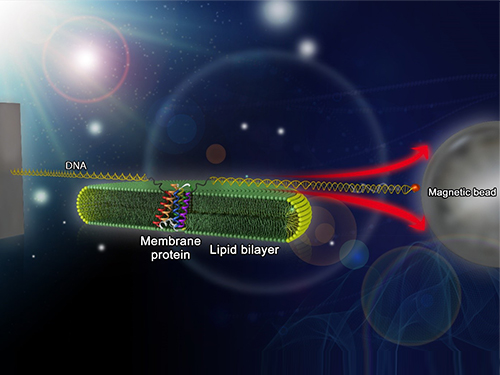 Mapping the Folding Process of a Single Membrane Protein
KAIST and UCLA scientists were able to observe an individual membrane protein fold and unfold by pulling and releasing magnetically trapped protein molecules.
Proteins are huge molecules containing hundreds to thousands of atoms that adopt a unique three dimensional structure, placing chemical groups in just the right place to catalyze reactions or build cellular structures. How all those atoms manage to find the right location - the so-called folding problem - has fascinated molecular biologists since the first structures were seen in the 1950s. Moreover, folding has important medical implications because most genetic defects cause protein misfolding.
About a third of all proteins float around in the cell membrane where they ensure the right chemicals get in the cell in the right amounts. Membrane proteins also provide key information links between the cell and its environment. Indeed, most drugs target membrane proteins. Nevertheless, the folding of membrane proteins has been particularly difficult to study and has rarely been studied in natural environments, leaving the folding process for a large fraction of the protein universe still largely cloaked in mystery.
In a recent issue of Nature Chemical Biology, published on October 19, 2015, a research team led by Tae-Young Yoon of the Department of Physics at the Korea Advanced Institute of Science and Technology (KAIST) and James U. Bowie of the Department of Chemistry and Biochemistry at the University of California, Los Angeles (UCLA), report a new method for manipulating the folding of membrane proteins in a membrane environment using a tool called a magnetic tweezer.
Researchers first attach long DNA handles to the ends of the protein. One handle is attached to a glass surface and the other to a magnetic bead. Using a magnet, they can essentially grab the protein and pull on it, inducing it to unfold. By playing with the bead attached to the protein, they can force the protein to unfold or allow it to refold, and watch all this happening by 3D-tracking of the magnetic bead. With this novel strategy, they were able to quantitatively map the folding energy landscape, the folding kinetic rate, and folding intermediates of a membrane protein in a membrane environment for the first time.
“I have been dreaming about this experiment for a decade. To see it work so well is really gratifying,” said Dr. Bowie.
One of the major surprises in the study was that essentially all the atoms of the protein jump into the correct structure together. The researchers expected that the protein structure would come together in a more piecemeal fashion, with different parts of the structure forming separately, but that was not the case. It is possible that nature evolved such a smooth, highly cooperative folding process to prevent partially folded forms that could get into trouble in the crowded cell membrane. On the other hand, the cooperative folding seen here might not apply to other membrane proteins.
“We need to look at more proteins. The technique developed here may allow us to do just that,” said Dr. Yoon.
The single molecule mechanical manipulation technique could enable detailed folding studies of many other membrane proteins. A major barrier to the study of membrane proteins previously is that the proteins tend to stick together and get tangled up, as computer cords lying at your feet tend to do. With the tweezer technique used in this work, the protein cords are held apart from other cords so they can’t get knotted up. It is hoped that the new approach will open up an important part of the protein universe to scrutiny, including many proteins that become misfolded in disease states.
The title of the research paper is “Mapping the energy landscape for second-stage folding of a single membrane protein” (DOI: 10.1038/nchembio.1939).
Picture: Single-molecule magnetic tweezers that induce mechanical unfolding and refolding of a single membrane protein. Since the force applied is parallel to the biological lipid membrane, the unfolding and refolding processes occur within the membrane.
2015.10.20 View 10257
Mapping the Folding Process of a Single Membrane Protein
KAIST and UCLA scientists were able to observe an individual membrane protein fold and unfold by pulling and releasing magnetically trapped protein molecules.
Proteins are huge molecules containing hundreds to thousands of atoms that adopt a unique three dimensional structure, placing chemical groups in just the right place to catalyze reactions or build cellular structures. How all those atoms manage to find the right location - the so-called folding problem - has fascinated molecular biologists since the first structures were seen in the 1950s. Moreover, folding has important medical implications because most genetic defects cause protein misfolding.
About a third of all proteins float around in the cell membrane where they ensure the right chemicals get in the cell in the right amounts. Membrane proteins also provide key information links between the cell and its environment. Indeed, most drugs target membrane proteins. Nevertheless, the folding of membrane proteins has been particularly difficult to study and has rarely been studied in natural environments, leaving the folding process for a large fraction of the protein universe still largely cloaked in mystery.
In a recent issue of Nature Chemical Biology, published on October 19, 2015, a research team led by Tae-Young Yoon of the Department of Physics at the Korea Advanced Institute of Science and Technology (KAIST) and James U. Bowie of the Department of Chemistry and Biochemistry at the University of California, Los Angeles (UCLA), report a new method for manipulating the folding of membrane proteins in a membrane environment using a tool called a magnetic tweezer.
Researchers first attach long DNA handles to the ends of the protein. One handle is attached to a glass surface and the other to a magnetic bead. Using a magnet, they can essentially grab the protein and pull on it, inducing it to unfold. By playing with the bead attached to the protein, they can force the protein to unfold or allow it to refold, and watch all this happening by 3D-tracking of the magnetic bead. With this novel strategy, they were able to quantitatively map the folding energy landscape, the folding kinetic rate, and folding intermediates of a membrane protein in a membrane environment for the first time.
“I have been dreaming about this experiment for a decade. To see it work so well is really gratifying,” said Dr. Bowie.
One of the major surprises in the study was that essentially all the atoms of the protein jump into the correct structure together. The researchers expected that the protein structure would come together in a more piecemeal fashion, with different parts of the structure forming separately, but that was not the case. It is possible that nature evolved such a smooth, highly cooperative folding process to prevent partially folded forms that could get into trouble in the crowded cell membrane. On the other hand, the cooperative folding seen here might not apply to other membrane proteins.
“We need to look at more proteins. The technique developed here may allow us to do just that,” said Dr. Yoon.
The single molecule mechanical manipulation technique could enable detailed folding studies of many other membrane proteins. A major barrier to the study of membrane proteins previously is that the proteins tend to stick together and get tangled up, as computer cords lying at your feet tend to do. With the tweezer technique used in this work, the protein cords are held apart from other cords so they can’t get knotted up. It is hoped that the new approach will open up an important part of the protein universe to scrutiny, including many proteins that become misfolded in disease states.
The title of the research paper is “Mapping the energy landscape for second-stage folding of a single membrane protein” (DOI: 10.1038/nchembio.1939).
Picture: Single-molecule magnetic tweezers that induce mechanical unfolding and refolding of a single membrane protein. Since the force applied is parallel to the biological lipid membrane, the unfolding and refolding processes occur within the membrane.
2015.10.20 View 10257 -
 KAIST holds the 2015 KAIST-MIT-Technion International Symposium on Nano Science
The 2015 KAIST-MIT-Technion International Symposium on Nano Science was held on August 11, 2015 at the KAIST campus. The event took place under three subtopics: Materials for Production and Storage of Renewable Energy, Functional Materials, and Multiferroic Materials.
The joint symposium invited more than 300 experts in material science and engineering including ten speakers and panelists. From MIT (Massachusetts Institute of Technology), Professors Harry L. Tuller and Geoffrey S. Beach in the Department of Material Engineering, as well as Professor Gregory Rutledge in the Department of Chemical Engineering joined the symposium. Professor Avner Rothschild in the Department of Material Engineering and Professor Yair Ein-Eli in the Department of Chemical Engineering at Technion Institute of Technology in Israel also participated.
From KAIST, Professors Il-Doo Kim, Byong-Guk Park, and Yeon-Sik Jung in the Department of Materials Science and Engineering, Professor Chan-Ho Yang in the Department of Physics, and Professor Doh-Chang Lee in the Department of Chemical and Biomolecular Engineering spoke at the event. The list of topics included “Next Generation Lithium-Air Battery,” “Nano Materials for High Performance Energy Storage System,” and “Iron Oxide Nanoparticles Photoelectrode for the Conversion and Storage of Solar Energy.”
In its efforts to promote cooperation among the three universities, KAIST plans to send six students to MIT and one student to Technion for joint research.
Professor Il-Doo Kim, who organized the symposium said, “We believe this kind of international gathering will serve as an opportunity for scholars from leading universities to share their expertise in material science and help them better understand on the recent trends in nanoscience and its related technology.”
2015.08.12 View 9038
KAIST holds the 2015 KAIST-MIT-Technion International Symposium on Nano Science
The 2015 KAIST-MIT-Technion International Symposium on Nano Science was held on August 11, 2015 at the KAIST campus. The event took place under three subtopics: Materials for Production and Storage of Renewable Energy, Functional Materials, and Multiferroic Materials.
The joint symposium invited more than 300 experts in material science and engineering including ten speakers and panelists. From MIT (Massachusetts Institute of Technology), Professors Harry L. Tuller and Geoffrey S. Beach in the Department of Material Engineering, as well as Professor Gregory Rutledge in the Department of Chemical Engineering joined the symposium. Professor Avner Rothschild in the Department of Material Engineering and Professor Yair Ein-Eli in the Department of Chemical Engineering at Technion Institute of Technology in Israel also participated.
From KAIST, Professors Il-Doo Kim, Byong-Guk Park, and Yeon-Sik Jung in the Department of Materials Science and Engineering, Professor Chan-Ho Yang in the Department of Physics, and Professor Doh-Chang Lee in the Department of Chemical and Biomolecular Engineering spoke at the event. The list of topics included “Next Generation Lithium-Air Battery,” “Nano Materials for High Performance Energy Storage System,” and “Iron Oxide Nanoparticles Photoelectrode for the Conversion and Storage of Solar Energy.”
In its efforts to promote cooperation among the three universities, KAIST plans to send six students to MIT and one student to Technion for joint research.
Professor Il-Doo Kim, who organized the symposium said, “We believe this kind of international gathering will serve as an opportunity for scholars from leading universities to share their expertise in material science and help them better understand on the recent trends in nanoscience and its related technology.”
2015.08.12 View 9038