%42%72%61%69%6e
-
 KAIST Develops mRNA Platform That Remains Effective Even in Aging and Obesity
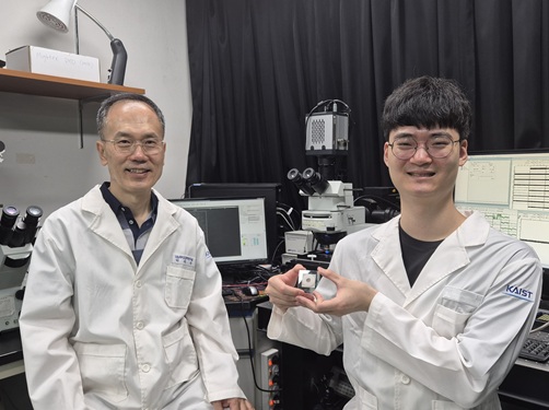
<(From Left) Dr. Subin Yoon, Ph.D candidate Hyeonggon Cho, Prof. Jae-Hwan Nam, Prof. Young-suk Lee>
Since the COVID-19 pandemic, mRNA vaccines have gained attention as a next-generation pharmaceutical technology. mRNA therapeutics work by delivering genetic instructions that enable cells to produce specific proteins for therapeutic effects. However, their efficacy has been reported to decline in elderly individuals or patients with obesity. To address this limitation, Korean researchers have newly designed a key regulatory region of mRNA that improves therapeutic protein production efficiency, developing a next-generation mRNA platform that maintains effectiveness even in aging and obesity conditions.
KAIST (President Kwang Hyung Lee) announced on the 10th of March that a joint research team led by Professor Young-suk Lee of the Department of Bio and Brain Engineering and Professor Jae-Hwan Nam of The Catholic University of Korea (President Jun-Gyu Choi) has developed a new mRNA platform by precisely designing the sequence of the 5′ untranslated region (5′UTR)*, a key regulatory region of mRNA.*5′ untranslated region (5′UTR): A region of mRNA that initiates and regulates protein production. The design of this region influences both the amount and speed of protein synthesis.
The research team analyzed large-scale bioinformatics datasets to identify 5′UTR sequences that enable proteins to be produced more efficiently across diverse cellular environments. When applied, the designed sequences significantly enhanced protein production and immune responses even in preclinical models of aging and obesity.
mRNA is a long single-stranded RNA molecule that serves as the blueprint for producing proteins required by the body. It consists of several components: the 5′UTR, which initiates and regulates the rate of protein production; the coding sequence (CDS), which contains the genetic information for a specific protein; the 3′ untranslated region (3′UTR), which helps maintain mRNA stability within cells; and the poly(A) tail, which further enhances stability and supports protein synthesis.
Among these components, the 5′UTR and 3′UTR do not determine the type of protein produced, but they play a critical role in regulating how efficiently the protein is synthesized. For this reason, these regions are receiving increasing attention as key bioengineering platforms for improving the performance of various mRNA therapeutics, including vaccines and treatments.
<Schematic Diagram of mRNA Therapeutic Design and Validation Using Bioinformatics>
To identify highly efficient 5′UTR sequences capable of promoting protein production across multiple tissues and cellular environments, the team conducted an integrated analysis of large-scale biological datasets. This included multiple analytical approaches such as RNA sequencing (RNA-seq) for analyzing gene activity across tissues, single-cell RNA sequencing (scRNA-seq) for examining gene expression at the individual cell level, and ribosome profiling (Ribo-seq) for measuring actual protein translation efficiency.
The researchers also focused on the fact that in aging or obesity conditions, cells often experience high levels of stress—particularly oxidative stress—which can reduce their ability to synthesize proteins. When the newly designed mRNA therapeutics were applied to preclinical models of aging and obesity, the results showed significantly improved protein production and immune responses compared with existing approaches. This research is expected to be applicable not only to mRNA vaccines but also to a wide range of biopharmaceutical technologies, including gene therapies and immunotherapies.
<Multimodal Bio–Big Data Analysis–Based mRNA Therapeutic Design (AI-Generated Image)>
Professor Young-suk Lee of KAIST Department of Bio and Brain Engineering stated, “This study identified a design strategy that enables mRNA to produce proteins more efficiently by analyzing large-scale biological data,” adding, “This technology will provide an important foundation for ensuring that mRNA vaccines and therapeutics remain effective even in environments where drug efficacy may decline, such as in elderly or obese patients.”
In this study, Dr. Subin Yoon from The Catholic University of Korea and doctoral candidate Hyeonggon Cho from KAIST participated as co-first authors. The research findings were published online on January 2 in the internationally renowned journal Molecular Therapy (IF = 12.0), a leading journal in gene and cell therapy.
(Paper title: ”Designing 5′UTR sequences improves the capacity of mRNA therapeutics in preclinical models of aging and obesity” DOI: https://doi.org/10.1016/j.ymthe.2025.12.060)
This research was supported by the Excellent Young Researcher Program and the Bio-Medical Technology Development Program of the National Research Foundation of Korea funded by the Ministry of Science and ICT, the Infectious Disease Response Innovative Technology Support Program of the Ministry of Food and Drug Safety, and the Infectious Disease Prevention and Therapeutics Technology Development Program of the Korea Health Industry Development Institute.
2026.03.10 View 1362
KAIST Develops mRNA Platform That Remains Effective Even in Aging and Obesity
<(From Left) Dr. Subin Yoon, Ph.D candidate Hyeonggon Cho, Prof. Jae-Hwan Nam, Prof. Young-suk Lee>
Since the COVID-19 pandemic, mRNA vaccines have gained attention as a next-generation pharmaceutical technology. mRNA therapeutics work by delivering genetic instructions that enable cells to produce specific proteins for therapeutic effects. However, their efficacy has been reported to decline in elderly individuals or patients with obesity. To address this limitation, Korean researchers have newly designed a key regulatory region of mRNA that improves therapeutic protein production efficiency, developing a next-generation mRNA platform that maintains effectiveness even in aging and obesity conditions.
KAIST (President Kwang Hyung Lee) announced on the 10th of March that a joint research team led by Professor Young-suk Lee of the Department of Bio and Brain Engineering and Professor Jae-Hwan Nam of The Catholic University of Korea (President Jun-Gyu Choi) has developed a new mRNA platform by precisely designing the sequence of the 5′ untranslated region (5′UTR)*, a key regulatory region of mRNA.*5′ untranslated region (5′UTR): A region of mRNA that initiates and regulates protein production. The design of this region influences both the amount and speed of protein synthesis.
The research team analyzed large-scale bioinformatics datasets to identify 5′UTR sequences that enable proteins to be produced more efficiently across diverse cellular environments. When applied, the designed sequences significantly enhanced protein production and immune responses even in preclinical models of aging and obesity.
mRNA is a long single-stranded RNA molecule that serves as the blueprint for producing proteins required by the body. It consists of several components: the 5′UTR, which initiates and regulates the rate of protein production; the coding sequence (CDS), which contains the genetic information for a specific protein; the 3′ untranslated region (3′UTR), which helps maintain mRNA stability within cells; and the poly(A) tail, which further enhances stability and supports protein synthesis.
Among these components, the 5′UTR and 3′UTR do not determine the type of protein produced, but they play a critical role in regulating how efficiently the protein is synthesized. For this reason, these regions are receiving increasing attention as key bioengineering platforms for improving the performance of various mRNA therapeutics, including vaccines and treatments.
<Schematic Diagram of mRNA Therapeutic Design and Validation Using Bioinformatics>
To identify highly efficient 5′UTR sequences capable of promoting protein production across multiple tissues and cellular environments, the team conducted an integrated analysis of large-scale biological datasets. This included multiple analytical approaches such as RNA sequencing (RNA-seq) for analyzing gene activity across tissues, single-cell RNA sequencing (scRNA-seq) for examining gene expression at the individual cell level, and ribosome profiling (Ribo-seq) for measuring actual protein translation efficiency.
The researchers also focused on the fact that in aging or obesity conditions, cells often experience high levels of stress—particularly oxidative stress—which can reduce their ability to synthesize proteins. When the newly designed mRNA therapeutics were applied to preclinical models of aging and obesity, the results showed significantly improved protein production and immune responses compared with existing approaches. This research is expected to be applicable not only to mRNA vaccines but also to a wide range of biopharmaceutical technologies, including gene therapies and immunotherapies.
<Multimodal Bio–Big Data Analysis–Based mRNA Therapeutic Design (AI-Generated Image)>
Professor Young-suk Lee of KAIST Department of Bio and Brain Engineering stated, “This study identified a design strategy that enables mRNA to produce proteins more efficiently by analyzing large-scale biological data,” adding, “This technology will provide an important foundation for ensuring that mRNA vaccines and therapeutics remain effective even in environments where drug efficacy may decline, such as in elderly or obese patients.”
In this study, Dr. Subin Yoon from The Catholic University of Korea and doctoral candidate Hyeonggon Cho from KAIST participated as co-first authors. The research findings were published online on January 2 in the internationally renowned journal Molecular Therapy (IF = 12.0), a leading journal in gene and cell therapy.
(Paper title: ”Designing 5′UTR sequences improves the capacity of mRNA therapeutics in preclinical models of aging and obesity” DOI: https://doi.org/10.1016/j.ymthe.2025.12.060)
This research was supported by the Excellent Young Researcher Program and the Bio-Medical Technology Development Program of the National Research Foundation of Korea funded by the Ministry of Science and ICT, the Infectious Disease Response Innovative Technology Support Program of the Ministry of Food and Drug Safety, and the Infectious Disease Prevention and Therapeutics Technology Development Program of the Korea Health Industry Development Institute.
2026.03.10 View 1362 -
 KAIST Develops Brain-Like AI… Thinks One More Time Even When Predictions Are Wrong
<(From left) Professor Sang Wan Lee, Myoung Hoon Ha, and Dr. Yoondo Sung>
Artificial intelligence now plays Go, paints pictures, and even converses like a human. However, there remains a decisive difference: AI requires far more electricity than the human brain to operate. Scientists have long asked the question, “How can the brain learn so intelligently using so little energy?” KAIST researchers have moved one step closer to the answer.
KAIST (President Kwang Hyung Lee) announced on the 29th that a research team led by Distinguished Professor Sang Wan Lee of the Department of Brain and Cognitive Sciences has developed a new technology that applies the learning principles of the human brain to deep learning, enabling stable training even in deep artificial intelligence models.
Our brain does not passively receive the world. Instead of merely perceiving what is happening in the present, it first predicts what will happen next and, when reality differs from that prediction, adjusts itself to reduce the difference (i.e., prediction error). This is similar to anticipating an opponent’s next move in Go and changing strategy if the prediction turns out to be wrong. This mode of information processing is known as “Predictive Coding.”
< Predictive Coding (PC) Module >
Scientists have attempted to apply this principle to AI, but encountered difficulties. As neural networks become deeper, errors tend to concentrate in specific layers or vanish altogether, repeatedly leading to performance degradation.
The research team mathematically identified the cause of this problem and proposed a new solution. The key idea is simple: instead of predicting only the final outcome, the AI is designed to also predict how its prediction errors will change in the future. The team refers to this as “Meta Prediction.” In simple terms, it is an AI that “thinks once more about its mistakes.” When this method was applied, learning proceeded stably in deep neural networks without halting.
<Analysis of Instability in Predictive Coding Model Errors>
The experimental results were also impressive. In 29 out of 30 experiments, the proposed method achieved higher accuracy than the current standard AI training method, backpropagation. Backpropagation is the representative learning method in which AI “goes backward by the amount of error and corrects it.”
Conventional AI training methods (backpropagation) require tightly interconnected layers, meaning the entire network must be computed and updated simultaneously. In contrast, this new approach demonstrates that, like the brain, large AI models can be effectively trained even when learning occurs in a distributed and partially independent manner.
<Performance Comparison of Predictive Coding Models>
This technology is expected to expand into various fields where power efficiency is critical, including neuromorphic computing, robot AI that must adapt to changing environments, and edge AI operating within devices.
Distinguished Professor Sang Wan Lee stated, “The key to this research is not simply imitating the structure of the brain, but enabling AI to follow the brain’s learning principles themselves,” adding, “We have opened the possibility of artificial intelligence that learns efficiently like the brain.”
This study was conducted with Dr. Myoung Hoon Ha as the first author and Professor Sang Wan Lee as the corresponding author. The paper was accepted to the International Conference on Learning Representations (ICLR 2026) and was published online on January 26.
※ Paper title: “Stable and Scalable Deep Predictive Coding Networks with Meta Prediction Errors”Original paper: https://openreview.net/forum?id=kE5jJUHl9i¬eId=e6T5T9cYqO
This research was supported by the Ministry of Science and ICT and the Institute of Information & Communications Technology Planning & Evaluation (IITP) through the Digital Global Research Support Program (joint research with Microsoft Research), the Samsung Electronics SAIT NPRC Program, and the SW Star Lab Program.
2026.03.06 View 1761
KAIST Develops Brain-Like AI… Thinks One More Time Even When Predictions Are Wrong
<(From left) Professor Sang Wan Lee, Myoung Hoon Ha, and Dr. Yoondo Sung>
Artificial intelligence now plays Go, paints pictures, and even converses like a human. However, there remains a decisive difference: AI requires far more electricity than the human brain to operate. Scientists have long asked the question, “How can the brain learn so intelligently using so little energy?” KAIST researchers have moved one step closer to the answer.
KAIST (President Kwang Hyung Lee) announced on the 29th that a research team led by Distinguished Professor Sang Wan Lee of the Department of Brain and Cognitive Sciences has developed a new technology that applies the learning principles of the human brain to deep learning, enabling stable training even in deep artificial intelligence models.
Our brain does not passively receive the world. Instead of merely perceiving what is happening in the present, it first predicts what will happen next and, when reality differs from that prediction, adjusts itself to reduce the difference (i.e., prediction error). This is similar to anticipating an opponent’s next move in Go and changing strategy if the prediction turns out to be wrong. This mode of information processing is known as “Predictive Coding.”
< Predictive Coding (PC) Module >
Scientists have attempted to apply this principle to AI, but encountered difficulties. As neural networks become deeper, errors tend to concentrate in specific layers or vanish altogether, repeatedly leading to performance degradation.
The research team mathematically identified the cause of this problem and proposed a new solution. The key idea is simple: instead of predicting only the final outcome, the AI is designed to also predict how its prediction errors will change in the future. The team refers to this as “Meta Prediction.” In simple terms, it is an AI that “thinks once more about its mistakes.” When this method was applied, learning proceeded stably in deep neural networks without halting.
<Analysis of Instability in Predictive Coding Model Errors>
The experimental results were also impressive. In 29 out of 30 experiments, the proposed method achieved higher accuracy than the current standard AI training method, backpropagation. Backpropagation is the representative learning method in which AI “goes backward by the amount of error and corrects it.”
Conventional AI training methods (backpropagation) require tightly interconnected layers, meaning the entire network must be computed and updated simultaneously. In contrast, this new approach demonstrates that, like the brain, large AI models can be effectively trained even when learning occurs in a distributed and partially independent manner.
<Performance Comparison of Predictive Coding Models>
This technology is expected to expand into various fields where power efficiency is critical, including neuromorphic computing, robot AI that must adapt to changing environments, and edge AI operating within devices.
Distinguished Professor Sang Wan Lee stated, “The key to this research is not simply imitating the structure of the brain, but enabling AI to follow the brain’s learning principles themselves,” adding, “We have opened the possibility of artificial intelligence that learns efficiently like the brain.”
This study was conducted with Dr. Myoung Hoon Ha as the first author and Professor Sang Wan Lee as the corresponding author. The paper was accepted to the International Conference on Learning Representations (ICLR 2026) and was published online on January 26.
※ Paper title: “Stable and Scalable Deep Predictive Coding Networks with Meta Prediction Errors”Original paper: https://openreview.net/forum?id=kE5jJUHl9i¬eId=e6T5T9cYqO
This research was supported by the Ministry of Science and ICT and the Institute of Information & Communications Technology Planning & Evaluation (IITP) through the Digital Global Research Support Program (joint research with Microsoft Research), the Samsung Electronics SAIT NPRC Program, and the SW Star Lab Program.
2026.03.06 View 1761 -
 Opening the Door to B Cell-Based Cancer-Remembering Personalized Cancer Vaccines
< (From left) KAIST Professor Jung Kyoon Choi, Dr. Jeong Yeon Kim, and Dr. Jin Hyeon An >
Neoantigens are unique markers that distinguish only cancer cells. By adding B cell reactivity, cancer vaccines can move beyond one-time attacks and short-term memory to become a long-term immunity that "remembers" cancer, effectively preventing recurrence. KAIST’s research team has developed an AI-based personalized cancer vaccine design technology that makes this possible and optimizes anticancer effects for each individual.
KAIST announced on January 2nd that Professor Jung Kyoon Choi’s research team from the Department of Bio and Brain Engineering, in collaboration with Neogen Logic Co., Ltd., has developed a new AI model to predict neoantigens—a core element of personalized cancer vaccine development—and clarified the importance of B cells in cancer immunotherapy.
The research team overcame the limitations of existing neoantigen discovery, which relied primarily on predicting T cell reactivity, and developed an AI-based neoantigen prediction technology that integrally considers both T cell and B cell reactivity.
This technology has been validated through large-scale cancer genome data, animal experiments, and clinical trial data for cancer vaccines. It is evaluated as the first AI technology capable of quantitatively predicting B cell reactivity to neoantigens.
Neoantigens are antigens composed of protein fragments derived from cancer cell mutations. Because they possess cancer-cell specificity, they have gained attention as a core target for next-generation cancer vaccines. Companies like Moderna and BioNTech developed COVID-19 vaccines using the mRNA platforms they secured while advancing neoantigen-based cancer vaccine technology, and they are currently actively conducting clinical trials for cancer vaccines alongside global pharmaceutical companies.
However, current cancer vaccine technology is mostly focused on T cell-centered immune responses, presenting a limitation in that it does not sufficiently reflect the immune responses mediated by B cells.
In fact, the research team of Professors Mark Yarchoan and Elizabeth Jaffee at Johns Hopkins University pointed out in Nature Reviews Cancer in May 2025 that “despite accumulating evidence regarding the role of B cells in tumor immunity, most cancer vaccine clinical trials still focus only on T cell responses.”
The research team’s new AI model overcomes existing limitations by learning the structural binding characteristics between mutant proteins and B cell receptors (BCR) to predict B cell reactivity. In particular, an analysis of cancer vaccine clinical trial data confirmed that integrating B cell responses can significantly enhance anti-tumor immune effects in actual clinical settings.
< Schematic Background of the Technology >
Professor Jung Kyoon Choi stated, “Together with Neogen Logic Co., Ltd., which is currently commercializing neoantigen AI technology, we are conducting pre-clinical development of a personalized cancer vaccine platform and are preparing to submit an FDA IND* with the goal of entering clinical trials in 2027.” He added, “We will enhance the scientific completeness of cancer vaccine development based on our proprietary AI technology and push forward the transition to the clinical stage step-by-step.”
*FDA IND: The procedure for obtaining permission from the U.S. Food and Drug Administration (FDA) to conduct clinical trials before administering a new drug to humans for the first time.
Dr. Jeong Yeon Kim and Dr. Jin Hyeon An participated as co-first authors in this study. The research results were published in the international scientific journal Science Advances on December 3rd.
※ Paper Title: B cell–reactive neoantigens boost antitumor immunity, DOI: 10.1126/sciadv.adx8303
2026.01.02 View 2646
Opening the Door to B Cell-Based Cancer-Remembering Personalized Cancer Vaccines
< (From left) KAIST Professor Jung Kyoon Choi, Dr. Jeong Yeon Kim, and Dr. Jin Hyeon An >
Neoantigens are unique markers that distinguish only cancer cells. By adding B cell reactivity, cancer vaccines can move beyond one-time attacks and short-term memory to become a long-term immunity that "remembers" cancer, effectively preventing recurrence. KAIST’s research team has developed an AI-based personalized cancer vaccine design technology that makes this possible and optimizes anticancer effects for each individual.
KAIST announced on January 2nd that Professor Jung Kyoon Choi’s research team from the Department of Bio and Brain Engineering, in collaboration with Neogen Logic Co., Ltd., has developed a new AI model to predict neoantigens—a core element of personalized cancer vaccine development—and clarified the importance of B cells in cancer immunotherapy.
The research team overcame the limitations of existing neoantigen discovery, which relied primarily on predicting T cell reactivity, and developed an AI-based neoantigen prediction technology that integrally considers both T cell and B cell reactivity.
This technology has been validated through large-scale cancer genome data, animal experiments, and clinical trial data for cancer vaccines. It is evaluated as the first AI technology capable of quantitatively predicting B cell reactivity to neoantigens.
Neoantigens are antigens composed of protein fragments derived from cancer cell mutations. Because they possess cancer-cell specificity, they have gained attention as a core target for next-generation cancer vaccines. Companies like Moderna and BioNTech developed COVID-19 vaccines using the mRNA platforms they secured while advancing neoantigen-based cancer vaccine technology, and they are currently actively conducting clinical trials for cancer vaccines alongside global pharmaceutical companies.
However, current cancer vaccine technology is mostly focused on T cell-centered immune responses, presenting a limitation in that it does not sufficiently reflect the immune responses mediated by B cells.
In fact, the research team of Professors Mark Yarchoan and Elizabeth Jaffee at Johns Hopkins University pointed out in Nature Reviews Cancer in May 2025 that “despite accumulating evidence regarding the role of B cells in tumor immunity, most cancer vaccine clinical trials still focus only on T cell responses.”
The research team’s new AI model overcomes existing limitations by learning the structural binding characteristics between mutant proteins and B cell receptors (BCR) to predict B cell reactivity. In particular, an analysis of cancer vaccine clinical trial data confirmed that integrating B cell responses can significantly enhance anti-tumor immune effects in actual clinical settings.
< Schematic Background of the Technology >
Professor Jung Kyoon Choi stated, “Together with Neogen Logic Co., Ltd., which is currently commercializing neoantigen AI technology, we are conducting pre-clinical development of a personalized cancer vaccine platform and are preparing to submit an FDA IND* with the goal of entering clinical trials in 2027.” He added, “We will enhance the scientific completeness of cancer vaccine development based on our proprietary AI technology and push forward the transition to the clinical stage step-by-step.”
*FDA IND: The procedure for obtaining permission from the U.S. Food and Drug Administration (FDA) to conduct clinical trials before administering a new drug to humans for the first time.
Dr. Jeong Yeon Kim and Dr. Jin Hyeon An participated as co-first authors in this study. The research results were published in the international scientific journal Science Advances on December 3rd.
※ Paper Title: B cell–reactive neoantigens boost antitumor immunity, DOI: 10.1126/sciadv.adx8303
2026.01.02 View 2646 -
 Thinking outside the box to Fabricate Customized 3D Neural Chips
<(From Left) Professor Yoonkey Nam, Dr. Dongjo Yoon from the Department of Bio and Brain Engineering>
Cultured neural tissues have been widely used as a simplified experimental model for brain research. However, existing devices for growing and recording neural tissues, which are manufactured using semiconductor processes, have limitations in terms of shape modification and the implementation of three-dimensional (3D) structures.
By "thinking outside the box," a KAIST research team has successfully created a customized 3D neural chip. They first used a 3D printer to fabricate a hollow channel structure, then used capillary action to automatically fill the channels with conductive ink, creating the electrodes and wiring. This achievement is expected to significantly increase the design freedom and versatility of brain science and brain engineering research platforms.
On the 25th, KAIST announced that a research team led by Professor Yoonkey Nam from the Department of Bio and Brain Engineering has successfully developed a platform technology that overcomes the limitations of traditional semiconductor-based manufacturing. This technology allows for the precise fabrication of "3D microelectrode array" (neural interfaces with multiple microelectrodes arranged in a 3D space to measure and stimulate the electrophysiological signal of neurons) in various customized forms for in vitro culture chips.
Existing 3D microelectrode array fabrication, based on semiconductor processes, has limited 3D design freedom and is expensive. While 3D printing-based fabrication techniques have recently been proposed to overcome these issues, they still have limitations in terms of 3D design freedom for various in vitro neural network structures because they follow the traditional sequence of "conductive material patterning → insulator coating → electrode opening."
The KAIST research team leveraged the excellent 3D design freedom provided by 3D printing technology and its ability to use printed materials as insulators. By reversing the traditional process, they established an innovative method that allows for more flexible design and functional measurement of 3D neuronal network models for in vitro culture.
<Schematic Diagram of an Integrated Cell Culture Substrate-Microelectrode Array Platform for In Vitro Cultured 3D Neural Network Models>
First, they used a 3D printer to print a hollow 3D insulator with micro-tunnels. This structure was designed to serve as a stable scaffold for conductive materials in 3D space while also supporting the creation of various 3D neuronal networks. They then demonstrated that by using capillary action to fill these internal micro-tunnels with conductive ink, they could create a 3D scaffold-microelectrode array with more freely arranged microelectrodes within a complex 3D culture support structure.
The new platform can be used to create various chip shapes, such as probe-type, cube-type, and modular-type, and supports the fabrication of electrodes using different materials like graphite, conductive polymers, and silver nanoparticles. This allows for the simultaneous measurement of multichannel neural signals from both inside and outside the 3D neuronal network, enabling precise analysis of the dynamic interactions and connectivity between neurons.
Professor Nam stated, "This research, which combines 3D printing and capillary action, is an achievement that significantly expands the freedom of neural chip fabrication." He added that it will contribute to the advancement of fundamental brain science research using neural tissue, as well as applied fields like cell-based biosensors and biocomputing.
Dr. Dongjo Yoon from KAIST's Department of Bio and Brain Engineering participated as the first author of the study. The research findings were published online in the international academic journal Advanced Functional Materials (June 25th issue).
※Paper Title: Highly Customizable Scaffold-Type 3D Microelectrode Array Platform for Design and Analysis of the 3D Neuronal Network In Vitro
This research was supported by the Consolidator Grants Program and the Global Basic Research Laboratory Program of the National Research Foundation of Korea.
2025.09.26 View 2659
Thinking outside the box to Fabricate Customized 3D Neural Chips
<(From Left) Professor Yoonkey Nam, Dr. Dongjo Yoon from the Department of Bio and Brain Engineering>
Cultured neural tissues have been widely used as a simplified experimental model for brain research. However, existing devices for growing and recording neural tissues, which are manufactured using semiconductor processes, have limitations in terms of shape modification and the implementation of three-dimensional (3D) structures.
By "thinking outside the box," a KAIST research team has successfully created a customized 3D neural chip. They first used a 3D printer to fabricate a hollow channel structure, then used capillary action to automatically fill the channels with conductive ink, creating the electrodes and wiring. This achievement is expected to significantly increase the design freedom and versatility of brain science and brain engineering research platforms.
On the 25th, KAIST announced that a research team led by Professor Yoonkey Nam from the Department of Bio and Brain Engineering has successfully developed a platform technology that overcomes the limitations of traditional semiconductor-based manufacturing. This technology allows for the precise fabrication of "3D microelectrode array" (neural interfaces with multiple microelectrodes arranged in a 3D space to measure and stimulate the electrophysiological signal of neurons) in various customized forms for in vitro culture chips.
Existing 3D microelectrode array fabrication, based on semiconductor processes, has limited 3D design freedom and is expensive. While 3D printing-based fabrication techniques have recently been proposed to overcome these issues, they still have limitations in terms of 3D design freedom for various in vitro neural network structures because they follow the traditional sequence of "conductive material patterning → insulator coating → electrode opening."
The KAIST research team leveraged the excellent 3D design freedom provided by 3D printing technology and its ability to use printed materials as insulators. By reversing the traditional process, they established an innovative method that allows for more flexible design and functional measurement of 3D neuronal network models for in vitro culture.
<Schematic Diagram of an Integrated Cell Culture Substrate-Microelectrode Array Platform for In Vitro Cultured 3D Neural Network Models>
First, they used a 3D printer to print a hollow 3D insulator with micro-tunnels. This structure was designed to serve as a stable scaffold for conductive materials in 3D space while also supporting the creation of various 3D neuronal networks. They then demonstrated that by using capillary action to fill these internal micro-tunnels with conductive ink, they could create a 3D scaffold-microelectrode array with more freely arranged microelectrodes within a complex 3D culture support structure.
The new platform can be used to create various chip shapes, such as probe-type, cube-type, and modular-type, and supports the fabrication of electrodes using different materials like graphite, conductive polymers, and silver nanoparticles. This allows for the simultaneous measurement of multichannel neural signals from both inside and outside the 3D neuronal network, enabling precise analysis of the dynamic interactions and connectivity between neurons.
Professor Nam stated, "This research, which combines 3D printing and capillary action, is an achievement that significantly expands the freedom of neural chip fabrication." He added that it will contribute to the advancement of fundamental brain science research using neural tissue, as well as applied fields like cell-based biosensors and biocomputing.
Dr. Dongjo Yoon from KAIST's Department of Bio and Brain Engineering participated as the first author of the study. The research findings were published online in the international academic journal Advanced Functional Materials (June 25th issue).
※Paper Title: Highly Customizable Scaffold-Type 3D Microelectrode Array Platform for Design and Analysis of the 3D Neuronal Network In Vitro
This research was supported by the Consolidator Grants Program and the Global Basic Research Laboratory Program of the National Research Foundation of Korea.
2025.09.26 View 2659 -
 The Secret of Our Success Author Joseph Henrich to Deliver Special Lecture at KAIST
KAIST announced on the 19th that its Institute for Mind and Brain Sciences and the Department of Brain and Cognitive Science will be hosting a special lecture by world-renowned cultural evolution scholar, Professor Joseph Henrich of Harvard University. The free lecture will take place on the 22nd at the Conference Room on the 1st floor of the Meta-Convergence Hall at the KAIST main campus, with support from the Gikwan Foundation. The event is open to the public.
Professor Henrich, a professor in the Department of Human Evolutionary Biology at Harvard, is a leading authority on the evolution of culture and cooperation. He was recognized for his work on the origins of human cooperative behavior through a comparative study of 15 small-scale societies, earning the 2024 Panmure House Prize* (Adam Smith 300th Anniversary Prize) and the 2022 Hayek Book Prize.
* Panmure House Prize: An academic award established in honor of Adam Smith's scholarship, named after the building where he lived.
< Poster for Special Lecture by Professor Joseph Henrich of Harvard University >
His representative books, "The WEIRDest People in the World" and "The Secret of Our Success," have created a significant stir in both academia and the general public by offering new interpretations of the formation and development of human society from a cultural evolution perspective.
"The WEIRDest People in the World" emphasizes that human thought and behavior are products of specific cultural environments rather than universal truths. "The Secret of Our Success" presents a new perspective on how humanity, through cultural artifacts like language, tools, and institutions, has achieved unique success compared to other animals.
The lecture will be divided into two sessions: an academic seminar and a public lecture. The academic seminar, held from 10:00 AM to 11:30 AM, will be conducted in English on the topic of "Cultural Evolutionary Psychology, Kinship, and the Historical Origins of Modern Psychological Differences." It is intended for researchers, graduate students, and undergraduate students in related fields.
Following this, a public lecture will be held from 3:00 PM to 5:00 PM on the topic of "The Collective Brain: Social and Cultural Origins of Creativity." Professor Jeong Jae-seung of KAIST's Department of Brain and Cognitive Science will serve as the moderator, and simultaneous interpretation will be provided.
The lecture will cover how innovation and creativity are products of a collective intelligence formed by diverse people exchanging ideas through networks. It will also discuss how the pace of innovation within a population is determined by key factors such as community size, social connectivity, and cognitive diversity, and how these principles explain innovation in various social contexts, including cultural psychology, immigration, urbanization, and institutions. There will also be a Q&A session with the author of "The Secret of Our Success."
Regarding the lecture, Professor Henrich stated, "In human evolution, culture is not just a backdrop; it's the core driving force that makes us human. Through this lecture, I want to share how we have learned from each other, cooperated, and developed knowledge and institutions. I especially look forward to having a deep conversation with the audience about the evolutionary significance of the passion for education and learning culture in Korean society."
Professor Jeong Jae-seung of KAIST's Department of Brain and Cognitive Science said, "This lecture was organized to explore how the human mind and brain have evolved through interaction with culture. It will be a valuable opportunity to hear the insights of a world-renowned scholar from the interdisciplinary perspective of meditation science and brain and cognitive science."
To register for the event, you can use the link (https://forms.gle/7TW9FAKv1qgA3dBBA) or the QR code on the poster. For inquiries, please contact the KAIST Institute for Mind and Brain Sciences at 042-350-1361.
2025.09.19 View 1955
The Secret of Our Success Author Joseph Henrich to Deliver Special Lecture at KAIST
KAIST announced on the 19th that its Institute for Mind and Brain Sciences and the Department of Brain and Cognitive Science will be hosting a special lecture by world-renowned cultural evolution scholar, Professor Joseph Henrich of Harvard University. The free lecture will take place on the 22nd at the Conference Room on the 1st floor of the Meta-Convergence Hall at the KAIST main campus, with support from the Gikwan Foundation. The event is open to the public.
Professor Henrich, a professor in the Department of Human Evolutionary Biology at Harvard, is a leading authority on the evolution of culture and cooperation. He was recognized for his work on the origins of human cooperative behavior through a comparative study of 15 small-scale societies, earning the 2024 Panmure House Prize* (Adam Smith 300th Anniversary Prize) and the 2022 Hayek Book Prize.
* Panmure House Prize: An academic award established in honor of Adam Smith's scholarship, named after the building where he lived.
< Poster for Special Lecture by Professor Joseph Henrich of Harvard University >
His representative books, "The WEIRDest People in the World" and "The Secret of Our Success," have created a significant stir in both academia and the general public by offering new interpretations of the formation and development of human society from a cultural evolution perspective.
"The WEIRDest People in the World" emphasizes that human thought and behavior are products of specific cultural environments rather than universal truths. "The Secret of Our Success" presents a new perspective on how humanity, through cultural artifacts like language, tools, and institutions, has achieved unique success compared to other animals.
The lecture will be divided into two sessions: an academic seminar and a public lecture. The academic seminar, held from 10:00 AM to 11:30 AM, will be conducted in English on the topic of "Cultural Evolutionary Psychology, Kinship, and the Historical Origins of Modern Psychological Differences." It is intended for researchers, graduate students, and undergraduate students in related fields.
Following this, a public lecture will be held from 3:00 PM to 5:00 PM on the topic of "The Collective Brain: Social and Cultural Origins of Creativity." Professor Jeong Jae-seung of KAIST's Department of Brain and Cognitive Science will serve as the moderator, and simultaneous interpretation will be provided.
The lecture will cover how innovation and creativity are products of a collective intelligence formed by diverse people exchanging ideas through networks. It will also discuss how the pace of innovation within a population is determined by key factors such as community size, social connectivity, and cognitive diversity, and how these principles explain innovation in various social contexts, including cultural psychology, immigration, urbanization, and institutions. There will also be a Q&A session with the author of "The Secret of Our Success."
Regarding the lecture, Professor Henrich stated, "In human evolution, culture is not just a backdrop; it's the core driving force that makes us human. Through this lecture, I want to share how we have learned from each other, cooperated, and developed knowledge and institutions. I especially look forward to having a deep conversation with the audience about the evolutionary significance of the passion for education and learning culture in Korean society."
Professor Jeong Jae-seung of KAIST's Department of Brain and Cognitive Science said, "This lecture was organized to explore how the human mind and brain have evolved through interaction with culture. It will be a valuable opportunity to hear the insights of a world-renowned scholar from the interdisciplinary perspective of meditation science and brain and cognitive science."
To register for the event, you can use the link (https://forms.gle/7TW9FAKv1qgA3dBBA) or the QR code on the poster. For inquiries, please contact the KAIST Institute for Mind and Brain Sciences at 042-350-1361.
2025.09.19 View 1955 -
 Accurate Real time ECG Measurement While Comfortably Lying Down at Home
< (From left) Professor Chul Kim of the Department of Bio and Brain Engineering, Ph.D. candidate Minjae Kim, researcher Premravee Teeravichayangoon >
KAIST's research team has developed a technology that can measure electrocardiogram (ECG) and heart rate variability (HRV) in real time by simply lying on a bed with clothes on, without having to go to the hospital. This technology is expected to evolve into a daily heart health monitoring platform in conjunction with remote healthcare, and further expand into various bio-healthcare fields such as sleep and stress analysis, contributing to personalized prevention and early diagnosis for patients.
KAIST announced on the 19th that Professor Chul Kim's research team from the Department of Bio and Brain Engineering has developed an 'in-bed cardiac monitoring on-device system'.
The research team manufactured a flexible substrate sensor that integrates the electronic circuit and electrodes into one to increase precision, and implemented an integrated system that can perform signal-noise separation, heart beat signal (R-peak) detection, and heart rate variability analysis in real time through on-device signal processing.
Existing ECG measurement had the inconvenience of visiting a hospital, taking off clothes, and attaching wet electrodes to the skin. Because of this, long-term monitoring was difficult, and it was not easy for the elderly or patients with chronic diseases to use it daily. Non-contact methods also had a technical limitation of being vulnerable to external noise.
To solve these problems, the research team applied a circuit that blocks external noise (active shielding) and a circuit that stably captures minute current changes in the human body (right-leg drive circuit). In addition, they implemented a mathematical transformation technique (wavelet transform) that extracts only the important parts from the heart beat signal and a calculation method (peak detection algorithm) that accurately identifies the moment of the heart's electrical beat (R-peak) as on-device signal processing techniques, allowing for precise real-time analysis of the signal.
As a result, users can obtain stable and accurate ECG signals even when lying on their backs with clothes on.
< Figure. Overall structural diagram of the developed non-contact in-bed cardiac monitoring on-device system, schematic diagram of the R-peak detection algorithm, real-time ECG and HRV measurement screen >
This research presents new possibilities for managing chronic cardiovascular diseases and supporting the health of the elderly, as it can be easily used not only in hospitals but also at home.
Professor Chul Kim said, "This system, which can extract signals in real time even in a noisy environment, can be used to easily check heart health in daily life," and added, "In the future, it will become the foundation of sleep health management by adding the measurement of various bio-signals."
This paper, in which Ph.D. candidate Minjae Kim and researcher Premravee Teeravichayangoon from the Department of Bio and Brain Engineering participated as co-first authors, was published online in the international journal 'Biosensors and Bioelectronics' on August 9, 2025.
※ Paper title: A homecare in-bed hardware system for precise real-time ECG and HRV monitoring with layered clothing. DOI: https://doi.org/10.1016/j.bios.2025.117838
※ Author information: Minjae Kim (KAIST Department of Bio and Brain Engineering, First Author), Premravee Teeravichayangoon (KAIST Department of Bio and Brain Engineering, First Author), Chul Kim (KAIST Department of Bio and Brain Engineering, Corresponding Author)
Meanwhile, this research was carried out with the support of the National Research Foundation of Korea's Basic Research Lab and Bio-medical Technology Development Project, and the KAIST-Ceragem Future Healthcare Research Center.
2025.09.19 View 1922
Accurate Real time ECG Measurement While Comfortably Lying Down at Home
< (From left) Professor Chul Kim of the Department of Bio and Brain Engineering, Ph.D. candidate Minjae Kim, researcher Premravee Teeravichayangoon >
KAIST's research team has developed a technology that can measure electrocardiogram (ECG) and heart rate variability (HRV) in real time by simply lying on a bed with clothes on, without having to go to the hospital. This technology is expected to evolve into a daily heart health monitoring platform in conjunction with remote healthcare, and further expand into various bio-healthcare fields such as sleep and stress analysis, contributing to personalized prevention and early diagnosis for patients.
KAIST announced on the 19th that Professor Chul Kim's research team from the Department of Bio and Brain Engineering has developed an 'in-bed cardiac monitoring on-device system'.
The research team manufactured a flexible substrate sensor that integrates the electronic circuit and electrodes into one to increase precision, and implemented an integrated system that can perform signal-noise separation, heart beat signal (R-peak) detection, and heart rate variability analysis in real time through on-device signal processing.
Existing ECG measurement had the inconvenience of visiting a hospital, taking off clothes, and attaching wet electrodes to the skin. Because of this, long-term monitoring was difficult, and it was not easy for the elderly or patients with chronic diseases to use it daily. Non-contact methods also had a technical limitation of being vulnerable to external noise.
To solve these problems, the research team applied a circuit that blocks external noise (active shielding) and a circuit that stably captures minute current changes in the human body (right-leg drive circuit). In addition, they implemented a mathematical transformation technique (wavelet transform) that extracts only the important parts from the heart beat signal and a calculation method (peak detection algorithm) that accurately identifies the moment of the heart's electrical beat (R-peak) as on-device signal processing techniques, allowing for precise real-time analysis of the signal.
As a result, users can obtain stable and accurate ECG signals even when lying on their backs with clothes on.
< Figure. Overall structural diagram of the developed non-contact in-bed cardiac monitoring on-device system, schematic diagram of the R-peak detection algorithm, real-time ECG and HRV measurement screen >
This research presents new possibilities for managing chronic cardiovascular diseases and supporting the health of the elderly, as it can be easily used not only in hospitals but also at home.
Professor Chul Kim said, "This system, which can extract signals in real time even in a noisy environment, can be used to easily check heart health in daily life," and added, "In the future, it will become the foundation of sleep health management by adding the measurement of various bio-signals."
This paper, in which Ph.D. candidate Minjae Kim and researcher Premravee Teeravichayangoon from the Department of Bio and Brain Engineering participated as co-first authors, was published online in the international journal 'Biosensors and Bioelectronics' on August 9, 2025.
※ Paper title: A homecare in-bed hardware system for precise real-time ECG and HRV monitoring with layered clothing. DOI: https://doi.org/10.1016/j.bios.2025.117838
※ Author information: Minjae Kim (KAIST Department of Bio and Brain Engineering, First Author), Premravee Teeravichayangoon (KAIST Department of Bio and Brain Engineering, First Author), Chul Kim (KAIST Department of Bio and Brain Engineering, Corresponding Author)
Meanwhile, this research was carried out with the support of the National Research Foundation of Korea's Basic Research Lab and Bio-medical Technology Development Project, and the KAIST-Ceragem Future Healthcare Research Center.
2025.09.19 View 1922 -
 KAIST Develops Smart Patch That Can Run Tests Using Sweat Instead of Blood
<(From Left) Ph.D candidate Jaehun Jeon, Professor Ki-Hun Jeong of the Department of Bio and Brain Engineering>
An era is opening where it's possible to precisely assess the body’s health status using only sweat instead of blood tests. A KAIST research team has developed a smart patch that can precisely observe internal changes through sweat when simply attached to the body. This is expected to greatly contribute to the advancement of chronic disease management and personalized healthcare technologies.
KAIST (President Kwang Hyung Lee) announced on September 7th that a research team led by Professor Ki-Hun Jeong of the Department of Bio and Brain Engineering has developed a wearable sensor that can simultaneously and in real-time analyze multiple metabolites in sweat.
Recently, research on wearable sensors that analyze metabolites in sweat to monitor the human body’s precise physiological state has been actively pursued. However, conventional “label-based” sensors, which require fluorescent tags or staining, and “label-free” methods have faced difficulties in effectively collecting and controlling sweat. Because of this, there have been limitations in precisely observing metabolite changes over time in actual human subjects.
<Figure 1. Flexible microfluidic nanoplasmonic patch (left). Sequential sample collection using the patch (center) and label-free metabolite profiling (right). In this study, we designed and fabricated a fully flexible nanoplasmonic microfluidic patch for label-free sweat analysis and performed SERS signal measurement and analysis directly from human sweat. Through this, we propose a platform capable of precisely identifying physiological changes induced by physical activity and dietary conditions.>
To overcome these limitations, the research team developed a thin and flexible wearable sweat patch that can be directly attached to the skin. This patch incorporates both microchannels for collecting sweat and an ultrafine nanoplasmonic structure* that label-freely analyzes sweat components using light. Thanks to this, multiple sweat metabolites can be simultaneously analyzed without the need for separate staining or labels, with just one patch application.
* Nanoplasmonic structure: An optical sensor structure where nanoscale metallic patterns interact with light, designed to sensitively detect the presence or changes in concentration of molecules in sweat.
The patch was created by combining nanophotonics technology, which manipulates light at the nanometer scale (one-hundred-thousandth the thickness of a human hair) to read molecular properties, with microfluidics technology, which precisely controls sweat in channels thinner than a hair.
In other words, within a single sweat patch, microfluidic technology enables sweat to be collected sequentially over time, allowing for the measurement of changes in various metabolites without any labeling process. Inside the patch are six to seventeen chambers (storage spaces), and sweat secreted during exercise flows along the microfluidic structures and fills each chamber in order.
<Figure 2. Example of the fabricated patch worn (left) and images of sequential sweat collection and storage (right). By designing precise microfluidic channels based on capillary burst valves, sequential sweat collection was implemented, which enabled label-free analysis of metabolite changes associated with exercise and diet.>
The research team applied the patch to actual human subjects and succeeded in continuously tracking the changing components of sweat over time during exercise. Previously, only about two components could be checked simultaneously through a label-free approach, but in this study, they demonstrated for the first time in the world that three metabolites—uric acid, lactic acid, and tyrosine—can be quantitatively analyzed simultaneously, as well as how they change depending on exercise and diet. In particular, by using artificial intelligence analysis methods, they were able to accurately distinguish signals of desired substances even within the complex components of sweat.
<Figure 3. Label-free analysis graphs of metabolite changes in sweat induced by exercise. Using the fabricated patch in combination with a machine learning model, metabolite concentrations in the sweat of actual subjects were analyzed. Comparison of sweat samples collected before and after consumption of a purine-rich diet, under exercise conditions, revealed label-free detection of changes in uric acid and tyrosine levels, as well as exercise-induced lactate increase. Validation experiments using commercial kits further confirmed the quantification accuracy, supporting the clinical applicability of this platform>
Professor Ki-Hun Jeong said, “This research lays the foundation for precisely monitoring internal metabolic changes over time without blood sampling by combining nanophotonics and microfluidics technologies.” He added, “In the future, it can be expanded to diverse fields such as chronic disease management, drug response tracking, environmental exposure monitoring, and the discovery of next-generation biomarkers for metabolic diseases.”
This research was conducted with Jaehun Jeon, a PhD student, as the first author and was published online in Nature Communications on August 27.
Paper Title: “All-Flexible Chronoepifluidic Nanoplasmonic Patch for Label-Free Metabolite Profiling in Sweat”
DOI: https://doi.org/10.1038/s41467-025-63510-2
This achievement was supported by the National Research Foundation of Korea, the Ministry of Science and ICT, the Ministry of Health and Welfare, and the Ministry of Trade, Industry and Energy.
2025.09.08 View 2522
KAIST Develops Smart Patch That Can Run Tests Using Sweat Instead of Blood
<(From Left) Ph.D candidate Jaehun Jeon, Professor Ki-Hun Jeong of the Department of Bio and Brain Engineering>
An era is opening where it's possible to precisely assess the body’s health status using only sweat instead of blood tests. A KAIST research team has developed a smart patch that can precisely observe internal changes through sweat when simply attached to the body. This is expected to greatly contribute to the advancement of chronic disease management and personalized healthcare technologies.
KAIST (President Kwang Hyung Lee) announced on September 7th that a research team led by Professor Ki-Hun Jeong of the Department of Bio and Brain Engineering has developed a wearable sensor that can simultaneously and in real-time analyze multiple metabolites in sweat.
Recently, research on wearable sensors that analyze metabolites in sweat to monitor the human body’s precise physiological state has been actively pursued. However, conventional “label-based” sensors, which require fluorescent tags or staining, and “label-free” methods have faced difficulties in effectively collecting and controlling sweat. Because of this, there have been limitations in precisely observing metabolite changes over time in actual human subjects.
<Figure 1. Flexible microfluidic nanoplasmonic patch (left). Sequential sample collection using the patch (center) and label-free metabolite profiling (right). In this study, we designed and fabricated a fully flexible nanoplasmonic microfluidic patch for label-free sweat analysis and performed SERS signal measurement and analysis directly from human sweat. Through this, we propose a platform capable of precisely identifying physiological changes induced by physical activity and dietary conditions.>
To overcome these limitations, the research team developed a thin and flexible wearable sweat patch that can be directly attached to the skin. This patch incorporates both microchannels for collecting sweat and an ultrafine nanoplasmonic structure* that label-freely analyzes sweat components using light. Thanks to this, multiple sweat metabolites can be simultaneously analyzed without the need for separate staining or labels, with just one patch application.
* Nanoplasmonic structure: An optical sensor structure where nanoscale metallic patterns interact with light, designed to sensitively detect the presence or changes in concentration of molecules in sweat.
The patch was created by combining nanophotonics technology, which manipulates light at the nanometer scale (one-hundred-thousandth the thickness of a human hair) to read molecular properties, with microfluidics technology, which precisely controls sweat in channels thinner than a hair.
In other words, within a single sweat patch, microfluidic technology enables sweat to be collected sequentially over time, allowing for the measurement of changes in various metabolites without any labeling process. Inside the patch are six to seventeen chambers (storage spaces), and sweat secreted during exercise flows along the microfluidic structures and fills each chamber in order.
<Figure 2. Example of the fabricated patch worn (left) and images of sequential sweat collection and storage (right). By designing precise microfluidic channels based on capillary burst valves, sequential sweat collection was implemented, which enabled label-free analysis of metabolite changes associated with exercise and diet.>
The research team applied the patch to actual human subjects and succeeded in continuously tracking the changing components of sweat over time during exercise. Previously, only about two components could be checked simultaneously through a label-free approach, but in this study, they demonstrated for the first time in the world that three metabolites—uric acid, lactic acid, and tyrosine—can be quantitatively analyzed simultaneously, as well as how they change depending on exercise and diet. In particular, by using artificial intelligence analysis methods, they were able to accurately distinguish signals of desired substances even within the complex components of sweat.
<Figure 3. Label-free analysis graphs of metabolite changes in sweat induced by exercise. Using the fabricated patch in combination with a machine learning model, metabolite concentrations in the sweat of actual subjects were analyzed. Comparison of sweat samples collected before and after consumption of a purine-rich diet, under exercise conditions, revealed label-free detection of changes in uric acid and tyrosine levels, as well as exercise-induced lactate increase. Validation experiments using commercial kits further confirmed the quantification accuracy, supporting the clinical applicability of this platform>
Professor Ki-Hun Jeong said, “This research lays the foundation for precisely monitoring internal metabolic changes over time without blood sampling by combining nanophotonics and microfluidics technologies.” He added, “In the future, it can be expanded to diverse fields such as chronic disease management, drug response tracking, environmental exposure monitoring, and the discovery of next-generation biomarkers for metabolic diseases.”
This research was conducted with Jaehun Jeon, a PhD student, as the first author and was published online in Nature Communications on August 27.
Paper Title: “All-Flexible Chronoepifluidic Nanoplasmonic Patch for Label-Free Metabolite Profiling in Sweat”
DOI: https://doi.org/10.1038/s41467-025-63510-2
This achievement was supported by the National Research Foundation of Korea, the Ministry of Science and ICT, the Ministry of Health and Welfare, and the Ministry of Trade, Industry and Energy.
2025.09.08 View 2522 -
 KAIST succeeds in controlling complex altered gene networks to restore them to normal
Previously, research on controlling gene networks has been carried out based on a single stimulus-response of cells. More recently, studies have been proposed to precisely analyze complex gene networks to identify control targets. A KAIST research team has succeeded in developing a universal technology that identifies gene control targets in altered cellular gene networks and restores them. This achievement is expected to be widely applied to new anticancer therapies such as cancer reversibility, drug development, precision medicine, and reprogramming for cell therapy.
KAIST (President Kwang Hyung Lee) announced on the 28th of August that Professor Kwang-Hyun Cho’s research team from the Department of Bio and Brain Engineering has developed a technology to systematically identify gene control targets that can restore the altered stimulus-response patterns of cells to normal by using an algebraic approach. The algebraic approach expresses gene networks as mathematical equations and identifies control targets through algebraic computations.
The research team represented the complex interactions among genes within a cell as a "logic circuit diagram" (Boolean network). Based on this, they visualized how a cell responds to external stimuli as a "landscape map" (phenotype landscape).
By applying a mathematical method called the "semi-tensor product,*" they developed a way to quickly and accurately calculate how the overall cellular response would change if a specific gene were controlled.
*Semi-tensor product: a method that calculates all possible gene combinations and control effects in a single algebraic formula
However, because the key genes that determine actual cellular responses number in the thousands, the calculations are extremely complex. To address this, the research team applied a numerical approximation method (Taylor approximation) to simplify the calculations. In simple terms, they transformed a complex problem into a simpler formula while still yielding nearly identical results.
Through this, the team was able to calculate which stable state (attractor) a cell would reach and predict how the cell’s state would change when a particular gene was controlled. As a result, they were able to identify core gene control targets that could restore abnormal cellular responses to states most similar to normal.
Professor Cho’s team applied the developed control technology to various gene networks and verified that it can accurately predict gene control targets that restore altered stimulus-response patterns of cells back to normal.
In particular, by applying it to bladder cancer cell networks, they identified gene control targets capable of restoring altered responses to normal. They also discovered gene control targets in large-scale distorted gene networks during immune cell differentiation that are capable of restoring normal stimulus-response patterns. This enabled them to solve problems that previously required only approximate searches through lengthy computer simulations in a fast and systematic way.
Professor Cho said, “This study is evaluated as a core original technology for the development of the Digital Cell Twin model*, which analyzes and controls the phenotype landscape of gene networks that determine cell fate. In the future, it is expected to be widely applicable across the life sciences and medicine, including new anticancer therapies through cancer reversibility, drug development, precision medicine, and reprogramming for cell therapy.”
*Digital Cell Twin model: a technology that digitally models the complex reactions occurring within cells, enabling virtual simulations of cellular responses instead of actual experiments
KAIST master’s student Insoo Jung, PhD student Corbin Hopper, PhD student Seong-Hoon Jang, and PhD student Hyunsoo Yeo participated in this study. The results were published online on August 22 in Science Advances, an international journal published by the American Association for the Advancement of Science (AAAS).
※ Paper title: “Reverse Control of Biological Networks to Restore Phenotype Landscapes”
※ DOI: https://www.science.org/doi/10.1126/sciadv.adw3995
This research was supported by the Mid-Career Researcher Program and the Basic Research Laboratory Program of the National Research Foundation of Korea, funded by the Ministry of Science and ICT.
2025.08.29 View 4532
KAIST succeeds in controlling complex altered gene networks to restore them to normal
Previously, research on controlling gene networks has been carried out based on a single stimulus-response of cells. More recently, studies have been proposed to precisely analyze complex gene networks to identify control targets. A KAIST research team has succeeded in developing a universal technology that identifies gene control targets in altered cellular gene networks and restores them. This achievement is expected to be widely applied to new anticancer therapies such as cancer reversibility, drug development, precision medicine, and reprogramming for cell therapy.
KAIST (President Kwang Hyung Lee) announced on the 28th of August that Professor Kwang-Hyun Cho’s research team from the Department of Bio and Brain Engineering has developed a technology to systematically identify gene control targets that can restore the altered stimulus-response patterns of cells to normal by using an algebraic approach. The algebraic approach expresses gene networks as mathematical equations and identifies control targets through algebraic computations.
The research team represented the complex interactions among genes within a cell as a "logic circuit diagram" (Boolean network). Based on this, they visualized how a cell responds to external stimuli as a "landscape map" (phenotype landscape).
By applying a mathematical method called the "semi-tensor product,*" they developed a way to quickly and accurately calculate how the overall cellular response would change if a specific gene were controlled.
*Semi-tensor product: a method that calculates all possible gene combinations and control effects in a single algebraic formula
However, because the key genes that determine actual cellular responses number in the thousands, the calculations are extremely complex. To address this, the research team applied a numerical approximation method (Taylor approximation) to simplify the calculations. In simple terms, they transformed a complex problem into a simpler formula while still yielding nearly identical results.
Through this, the team was able to calculate which stable state (attractor) a cell would reach and predict how the cell’s state would change when a particular gene was controlled. As a result, they were able to identify core gene control targets that could restore abnormal cellular responses to states most similar to normal.
Professor Cho’s team applied the developed control technology to various gene networks and verified that it can accurately predict gene control targets that restore altered stimulus-response patterns of cells back to normal.
In particular, by applying it to bladder cancer cell networks, they identified gene control targets capable of restoring altered responses to normal. They also discovered gene control targets in large-scale distorted gene networks during immune cell differentiation that are capable of restoring normal stimulus-response patterns. This enabled them to solve problems that previously required only approximate searches through lengthy computer simulations in a fast and systematic way.
Professor Cho said, “This study is evaluated as a core original technology for the development of the Digital Cell Twin model*, which analyzes and controls the phenotype landscape of gene networks that determine cell fate. In the future, it is expected to be widely applicable across the life sciences and medicine, including new anticancer therapies through cancer reversibility, drug development, precision medicine, and reprogramming for cell therapy.”
*Digital Cell Twin model: a technology that digitally models the complex reactions occurring within cells, enabling virtual simulations of cellular responses instead of actual experiments
KAIST master’s student Insoo Jung, PhD student Corbin Hopper, PhD student Seong-Hoon Jang, and PhD student Hyunsoo Yeo participated in this study. The results were published online on August 22 in Science Advances, an international journal published by the American Association for the Advancement of Science (AAAS).
※ Paper title: “Reverse Control of Biological Networks to Restore Phenotype Landscapes”
※ DOI: https://www.science.org/doi/10.1126/sciadv.adw3995
This research was supported by the Mid-Career Researcher Program and the Basic Research Laboratory Program of the National Research Foundation of Korea, funded by the Ministry of Science and ICT.
2025.08.29 View 4532 -
 Immune Signals Directly Modulate Brain's Emotional Circuits: Unraveling the Mechanism Behind Anxiety-Inducing Behaviors
KAIST's Department of Brain and Cognitive Sciences, led by Professor Jeong-Tae Kwon, has collaborated with MIT and Harvard Medical School to make a groundbreaking discovery. For the first time globally, their joint research has revealed that cytokines, released during immune responses, directly influence the brain's emotional circuits to regulate anxiety behavior.
The study provided experimental evidence for a bidirectional regulatory mechanism: inflammatory cytokines IL-17A and IL-17C act on specific neurons in the amygdala, a region known for emotional regulation, increasing their excitability and consequently inducing anxiety. Conversely, the anti-inflammatory cytokine IL-10 was found to suppress excitability in these very same neurons, thereby contributing to anxiety alleviation.
In a mouse model, the research team observed that while skin inflammation was mitigated by immunotherapy (IL-17RA antibody), anxiety levels paradoxically rose. This was attributed to elevated circulating IL-17 family cytokines leading to the overactivation of amygdala neurons.
Key finding: Inflammatory cytokines IL-17A/17C promote anxiety by acting on excitable amygdala neurons (via IL-17RA/RE receptors), whereas anti-inflammatory cytokine IL-10 alleviates anxiety by suppressing excitability through IL-10RA receptors on the same neurons.
The researchers further elucidated that the anti-inflammatory cytokine IL-10 works to reduce the excitability of these amygdala neurons, thereby mitigating anxiety responses.
This research marks the first instance of demonstrating that immune responses, such as infections or inflammation, directly impact emotional regulation at the level of brain circuits, extending beyond simple physical reactions. This is a profoundly significant achievement, as it proposes a crucial biological mechanism that interlinks immunity, emotion, and behavior through identical neurons within the brain.
The findings of this research were published in the esteemed international journal Cell on April 17th of this year.
Paper Information:
Title: Inflammatory and anti-inflammatory cytokines bidirectionally modulate amygdala circuits regulating anxiety
Journal: Cell (Vol. 188, 2190–2220), April 17, 2025
DOI: https://doi.org/10.1016/j.cell.2025.03.005
Corresponding Authors: Professor Gloria Choi (MIT), Professor Jun R. Huh (Harvard Medical School)
2025.07.24 View 3603
Immune Signals Directly Modulate Brain's Emotional Circuits: Unraveling the Mechanism Behind Anxiety-Inducing Behaviors
KAIST's Department of Brain and Cognitive Sciences, led by Professor Jeong-Tae Kwon, has collaborated with MIT and Harvard Medical School to make a groundbreaking discovery. For the first time globally, their joint research has revealed that cytokines, released during immune responses, directly influence the brain's emotional circuits to regulate anxiety behavior.
The study provided experimental evidence for a bidirectional regulatory mechanism: inflammatory cytokines IL-17A and IL-17C act on specific neurons in the amygdala, a region known for emotional regulation, increasing their excitability and consequently inducing anxiety. Conversely, the anti-inflammatory cytokine IL-10 was found to suppress excitability in these very same neurons, thereby contributing to anxiety alleviation.
In a mouse model, the research team observed that while skin inflammation was mitigated by immunotherapy (IL-17RA antibody), anxiety levels paradoxically rose. This was attributed to elevated circulating IL-17 family cytokines leading to the overactivation of amygdala neurons.
Key finding: Inflammatory cytokines IL-17A/17C promote anxiety by acting on excitable amygdala neurons (via IL-17RA/RE receptors), whereas anti-inflammatory cytokine IL-10 alleviates anxiety by suppressing excitability through IL-10RA receptors on the same neurons.
The researchers further elucidated that the anti-inflammatory cytokine IL-10 works to reduce the excitability of these amygdala neurons, thereby mitigating anxiety responses.
This research marks the first instance of demonstrating that immune responses, such as infections or inflammation, directly impact emotional regulation at the level of brain circuits, extending beyond simple physical reactions. This is a profoundly significant achievement, as it proposes a crucial biological mechanism that interlinks immunity, emotion, and behavior through identical neurons within the brain.
The findings of this research were published in the esteemed international journal Cell on April 17th of this year.
Paper Information:
Title: Inflammatory and anti-inflammatory cytokines bidirectionally modulate amygdala circuits regulating anxiety
Journal: Cell (Vol. 188, 2190–2220), April 17, 2025
DOI: https://doi.org/10.1016/j.cell.2025.03.005
Corresponding Authors: Professor Gloria Choi (MIT), Professor Jun R. Huh (Harvard Medical School)
2025.07.24 View 3603 -
 KAIST Successfully Implements 3D Brain-Mimicking Platform with 6x Higher Precision
<(From left) Dr. Dongjo Yoon, Professor Je-Kyun Park from the Department of Bio and Brain Engineering, (upper right) Professor Yoonkey Nam, Dr. Soo Jee Kim>
Existing three-dimensional (3D) neuronal culture technology has limitations in brain research due to the difficulty of precisely replicating the brain's complex multilayered structure and the lack of a platform that can simultaneously analyze both structure and function. A KAIST research team has successfully developed an integrated platform that can implement brain-like layered neuronal structures using 3D printing technology and precisely measure neuronal activity within them.
KAIST (President Kwang Hyung Lee) announced on the 16th of July that a joint research team led by Professors Je-Kyun Park and Yoonkey Nam from the Department of Bio and Brain Engineering has developed an integrated platform capable of fabricating high-resolution 3D multilayer neuronal networks using low-viscosity natural hydrogels with mechanical properties similar to brain tissue, and simultaneously analyzing their structural and functional connectivity.
Conventional bioprinting technology uses high-viscosity bioinks for structural stability, but this limits neuronal proliferation and neurite growth. Conversely, neural cell-friendly low-viscosity hydrogels are difficult to precisely pattern, leading to a fundamental trade-off between structural stability and biological function. The research team completed a sophisticated and stable brain-mimicking platform by combining three key technologies that enable the precise creation of brain structure with dilute gels, accurate alignment between layers, and simultaneous observation of neuronal activity.
The three core technologies are: ▲ 'Capillary Pinning Effect' technology, which enables the dilute gel (hydrogel) to adhere firmly to a stainless steel mesh (micromesh) to prevent it from flowing, thereby reproducing brain structures with six times greater precision (resolution of 500 μm or less) than conventional methods; ▲ the '3D Printing Aligner,' a cylindrical design that ensures the printed layers are precisely stacked without misalignment, guaranteeing the accurate assembly of multilayer structures and stable integration with microelectrode chips; and ▲ 'Dual-mode Analysis System' technology, which simultaneously measures electrical signals from below and observes cell activity with light (calcium imaging) from above, allowing for the simultaneous verification of the functional operation of interlayer connections through multiple methods.
< Figure 1. Platform integrating brain-structure-mimicking neural network model construction and functional measurement technology>
The research team successfully implemented a three-layered mini-brain structure using 3D printing with a fibrin hydrogel, which has elastic properties similar to those of the brain, and experimentally verified the process of actual neural cells transmitting and receiving signals within it.
Cortical neurons were placed in the upper and lower layers, while the middle layer was left empty but designed to allow neurons to penetrate and connect through it. Electrical signals were measured from the lower layer using a microsensor (electrode chip), and cell activity was observed from the upper layer using light (calcium imaging). The results showed that when electrical stimulation was applied, neural cells in both upper and lower layers responded simultaneously. When a synapse-blocking agent (synaptic blocker) was introduced, the response decreased, proving that the neural cells were genuinely connected and transmitting signals.
Professor Je-Kyun Park of KAIST explained, "This research is a joint development achievement of an integrated platform that can simultaneously reproduce the complex multilayered structure and function of brain tissue. Compared to existing technologies where signal measurement was impossible for more than 14 days, this platform maintains a stable microelectrode chip interface for over 27 days, allowing the real-time analysis of structure-function relationships. It can be utilized in various brain research fields such as neurological disease modeling, brain function research, neurotoxicity assessment, and neuroprotective drug screening in the future."
The research, in which Dr. Soo Jee Kim and Dr. Dongjo Yoon from KAIST's Department of Bio and Brain Engineering participated as co-first authors, was published online in the international journal 'Biosensors and Bioelectronics' on June 11, 2025.
※Paper: Hybrid biofabrication of multilayered 3D neuronal networks with structural and functional interlayer connectivity
※DOI: https://doi.org/10.1016/j.bios.2025.117688
2025.07.16 View 4335
KAIST Successfully Implements 3D Brain-Mimicking Platform with 6x Higher Precision
<(From left) Dr. Dongjo Yoon, Professor Je-Kyun Park from the Department of Bio and Brain Engineering, (upper right) Professor Yoonkey Nam, Dr. Soo Jee Kim>
Existing three-dimensional (3D) neuronal culture technology has limitations in brain research due to the difficulty of precisely replicating the brain's complex multilayered structure and the lack of a platform that can simultaneously analyze both structure and function. A KAIST research team has successfully developed an integrated platform that can implement brain-like layered neuronal structures using 3D printing technology and precisely measure neuronal activity within them.
KAIST (President Kwang Hyung Lee) announced on the 16th of July that a joint research team led by Professors Je-Kyun Park and Yoonkey Nam from the Department of Bio and Brain Engineering has developed an integrated platform capable of fabricating high-resolution 3D multilayer neuronal networks using low-viscosity natural hydrogels with mechanical properties similar to brain tissue, and simultaneously analyzing their structural and functional connectivity.
Conventional bioprinting technology uses high-viscosity bioinks for structural stability, but this limits neuronal proliferation and neurite growth. Conversely, neural cell-friendly low-viscosity hydrogels are difficult to precisely pattern, leading to a fundamental trade-off between structural stability and biological function. The research team completed a sophisticated and stable brain-mimicking platform by combining three key technologies that enable the precise creation of brain structure with dilute gels, accurate alignment between layers, and simultaneous observation of neuronal activity.
The three core technologies are: ▲ 'Capillary Pinning Effect' technology, which enables the dilute gel (hydrogel) to adhere firmly to a stainless steel mesh (micromesh) to prevent it from flowing, thereby reproducing brain structures with six times greater precision (resolution of 500 μm or less) than conventional methods; ▲ the '3D Printing Aligner,' a cylindrical design that ensures the printed layers are precisely stacked without misalignment, guaranteeing the accurate assembly of multilayer structures and stable integration with microelectrode chips; and ▲ 'Dual-mode Analysis System' technology, which simultaneously measures electrical signals from below and observes cell activity with light (calcium imaging) from above, allowing for the simultaneous verification of the functional operation of interlayer connections through multiple methods.
< Figure 1. Platform integrating brain-structure-mimicking neural network model construction and functional measurement technology>
The research team successfully implemented a three-layered mini-brain structure using 3D printing with a fibrin hydrogel, which has elastic properties similar to those of the brain, and experimentally verified the process of actual neural cells transmitting and receiving signals within it.
Cortical neurons were placed in the upper and lower layers, while the middle layer was left empty but designed to allow neurons to penetrate and connect through it. Electrical signals were measured from the lower layer using a microsensor (electrode chip), and cell activity was observed from the upper layer using light (calcium imaging). The results showed that when electrical stimulation was applied, neural cells in both upper and lower layers responded simultaneously. When a synapse-blocking agent (synaptic blocker) was introduced, the response decreased, proving that the neural cells were genuinely connected and transmitting signals.
Professor Je-Kyun Park of KAIST explained, "This research is a joint development achievement of an integrated platform that can simultaneously reproduce the complex multilayered structure and function of brain tissue. Compared to existing technologies where signal measurement was impossible for more than 14 days, this platform maintains a stable microelectrode chip interface for over 27 days, allowing the real-time analysis of structure-function relationships. It can be utilized in various brain research fields such as neurological disease modeling, brain function research, neurotoxicity assessment, and neuroprotective drug screening in the future."
The research, in which Dr. Soo Jee Kim and Dr. Dongjo Yoon from KAIST's Department of Bio and Brain Engineering participated as co-first authors, was published online in the international journal 'Biosensors and Bioelectronics' on June 11, 2025.
※Paper: Hybrid biofabrication of multilayered 3D neuronal networks with structural and functional interlayer connectivity
※DOI: https://doi.org/10.1016/j.bios.2025.117688
2025.07.16 View 4335 -
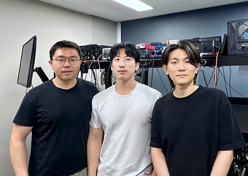 High-Resolution Spectrometer that Fits into Smartphones Developed by KAIST Researchers
- Professor Mooseok Jang's research team at the Department of Bio and Brain Engineering develops an ultra-compact, high-resolution spectrometer using 'double-layer disordered metasurfaces' that generate unique random patterns depending on light's color.
- Unlike conventional dispersion-based spectrometers that were difficult to apply to portable devices, this new concept spectrometer technology achieves 1nm-level high resolution in a device smaller than 1cm, comparable in size to a fingernail.
- It can be utilized as a built-in spectrometer in smartphones and wearable devices in the future, and can be expanded to advanced optical technologies such as hyperspectral imaging and ultrafast imaging.
< Photo 1. (From left) Professor Mooseok Jang, Dong-gu Lee (Ph.D. candidate), Gookho Song (Ph.D. candidate) >
Color, as the way light's wavelength is perceived by the human eye, goes beyond a simple aesthetic element, containing important scientific information like a substance's composition or state. Spectrometers are optical devices that analyze material properties by decomposing light into its constituent wavelengths, and they are widely used in various scientific and industrial fields, including material analysis, chemical component detection, and life science research. Existing high-resolution spectrometers were large and complex, making them difficult for widespread daily use. However, thanks to the ultra-compact, high-resolution spectrometer developed by KAIST researchers, it is now expected that light's color information can be utilized even within smartphones or wearable devices.
KAIST (President Kwang Hyung Lee) announced on the 13th that Professor Mooseok Jang's research team at the Department of Bio and Brain Engineering has successfully developed a reconstruction-based spectrometer technology using double-layer disordered metasurfaces*.
*Double-layer disordered metasurface: An innovative optical device that complexly scatters light through two layers of disordered nanostructures, creating unique and predictable speckle patterns for each wavelength.
Existing high-resolution spectrometers have a large form factor, on the order of tens of centimeters, and require complex calibration processes to maintain accuracy. This fundamentally stems from the operating principle of traditional dispersive elements, such as gratings and prisms, which separate light wavelengths along the propagation direction, much like a rainbow separates colors. Consequently, despite the potential for light's color information to be widely useful in daily life, spectroscopic technology has been limited to laboratory or industrial manufacturing environments.
< Figure 1. Through a simple structure consisting of a double layer of disordered metasurfaces and an image sensor, it was shown that speckles of predictable spectral channels with high spectral resolution can be generated in a compact form factor. The high similarity between the measured and calculated speckles was used to solve the inverse problem and verify the ability to reconstruct the spectrum. >
The research team devised a method that departs from the conventional spectroscopic paradigm of using diffraction gratings or prisms, which establish a one-to-one correspondence between light's color information and its propagation direction, by utilizing designed disordered structures as optical components. In this process, they employed metasurfaces, which can freely control the light propagation process using structures tens to hundreds of nanometers in size, to accurately implement 'complex random patterns (speckle*)'.
*Speckle: An irregular pattern of light intensity created by the interference of multiple wavefronts of light.
Specifically, they developed a method that involves implementing a double-layer disordered metasurface to generate wavelength-specific speckle patterns and then reconstructing precise color information (wavelength) of the light from the random patterns measured by a camera.
As a result, they successfully developed a new concept spectrometer technology that can accurately measure light across a broad range of visible to infrared (440-1,300nm) with a high resolution of 1 nanometer (nm) in a device smaller than a fingernail (less than 1cm) using only a single image capture.
< Figure 2. A disordered metasurface is a metasurface with irregularly arranged structures ranging from tens to hundreds of nanometers in size. In a double-layer structure, a propagation space is placed between the two metasurfaces to control the output speckle with high degrees of freedom, thereby achieving a spectral resolution of 1 nm even in a form factor smaller than 1 cm. >
Dong-gu Lee, a lead author of this study, stated, "This technology is implemented in a way that is directly integrated with commercial image sensors, and we expect that it will enable easy acquisition and utilization of light's wavelength information in daily life when built into mobile devices in the future."
Professor Mooseok Jang said, "This technology overcomes the limitations of existing RGB three-color based machine vision fields, which only distinguish and recognize three color components (red, green, blue), and has diverse applications. We anticipate various applied research for this technology, which expands the horizon of laboratory-level technology to daily-level machine vision technology for applications such as food component analysis, crop health diagnosis, skin health measurement, environmental pollution detection, and bio/medical diagnostics." He added, "Furthermore, it can be extended to various advanced optical technologies such as hyperspectral imaging, which records wavelength and spatial information simultaneously with high resolution, 3D optical trapping technology, which precisely controls light of multiple wavelengths into desired forms, and ultrafast imaging technology, which captures phenomena occurring in very short periods."
This research was collaboratively led by Dong-gu Lee (Ph.D. candidate) and Gookho Song (Ph.D. candidate) from the KAIST Department of Bio and Brain Engineering as co-first authors, with Professor Mooseok Jang as the corresponding author. The findings were published online in the international journal Science Advances on May 28, 2025.* Paper Title: Reconstructive spectrometer using double-layer disordered metasurfaces* DOI: 10.1126/sciadv.adv2376
This research was supported by the Samsung Research Funding and Incubation Center of Samsung Electronics grant, the National Research Foundation of Korea (NRF) grant funded by the Korea government (MSIT), and the Bio & Medical Technology Development Program of the National Research Foundation (NRF) funded by the Korean government (MSIT).
2025.06.13 View 8186
High-Resolution Spectrometer that Fits into Smartphones Developed by KAIST Researchers
- Professor Mooseok Jang's research team at the Department of Bio and Brain Engineering develops an ultra-compact, high-resolution spectrometer using 'double-layer disordered metasurfaces' that generate unique random patterns depending on light's color.
- Unlike conventional dispersion-based spectrometers that were difficult to apply to portable devices, this new concept spectrometer technology achieves 1nm-level high resolution in a device smaller than 1cm, comparable in size to a fingernail.
- It can be utilized as a built-in spectrometer in smartphones and wearable devices in the future, and can be expanded to advanced optical technologies such as hyperspectral imaging and ultrafast imaging.
< Photo 1. (From left) Professor Mooseok Jang, Dong-gu Lee (Ph.D. candidate), Gookho Song (Ph.D. candidate) >
Color, as the way light's wavelength is perceived by the human eye, goes beyond a simple aesthetic element, containing important scientific information like a substance's composition or state. Spectrometers are optical devices that analyze material properties by decomposing light into its constituent wavelengths, and they are widely used in various scientific and industrial fields, including material analysis, chemical component detection, and life science research. Existing high-resolution spectrometers were large and complex, making them difficult for widespread daily use. However, thanks to the ultra-compact, high-resolution spectrometer developed by KAIST researchers, it is now expected that light's color information can be utilized even within smartphones or wearable devices.
KAIST (President Kwang Hyung Lee) announced on the 13th that Professor Mooseok Jang's research team at the Department of Bio and Brain Engineering has successfully developed a reconstruction-based spectrometer technology using double-layer disordered metasurfaces*.
*Double-layer disordered metasurface: An innovative optical device that complexly scatters light through two layers of disordered nanostructures, creating unique and predictable speckle patterns for each wavelength.
Existing high-resolution spectrometers have a large form factor, on the order of tens of centimeters, and require complex calibration processes to maintain accuracy. This fundamentally stems from the operating principle of traditional dispersive elements, such as gratings and prisms, which separate light wavelengths along the propagation direction, much like a rainbow separates colors. Consequently, despite the potential for light's color information to be widely useful in daily life, spectroscopic technology has been limited to laboratory or industrial manufacturing environments.
< Figure 1. Through a simple structure consisting of a double layer of disordered metasurfaces and an image sensor, it was shown that speckles of predictable spectral channels with high spectral resolution can be generated in a compact form factor. The high similarity between the measured and calculated speckles was used to solve the inverse problem and verify the ability to reconstruct the spectrum. >
The research team devised a method that departs from the conventional spectroscopic paradigm of using diffraction gratings or prisms, which establish a one-to-one correspondence between light's color information and its propagation direction, by utilizing designed disordered structures as optical components. In this process, they employed metasurfaces, which can freely control the light propagation process using structures tens to hundreds of nanometers in size, to accurately implement 'complex random patterns (speckle*)'.
*Speckle: An irregular pattern of light intensity created by the interference of multiple wavefronts of light.
Specifically, they developed a method that involves implementing a double-layer disordered metasurface to generate wavelength-specific speckle patterns and then reconstructing precise color information (wavelength) of the light from the random patterns measured by a camera.
As a result, they successfully developed a new concept spectrometer technology that can accurately measure light across a broad range of visible to infrared (440-1,300nm) with a high resolution of 1 nanometer (nm) in a device smaller than a fingernail (less than 1cm) using only a single image capture.
< Figure 2. A disordered metasurface is a metasurface with irregularly arranged structures ranging from tens to hundreds of nanometers in size. In a double-layer structure, a propagation space is placed between the two metasurfaces to control the output speckle with high degrees of freedom, thereby achieving a spectral resolution of 1 nm even in a form factor smaller than 1 cm. >
Dong-gu Lee, a lead author of this study, stated, "This technology is implemented in a way that is directly integrated with commercial image sensors, and we expect that it will enable easy acquisition and utilization of light's wavelength information in daily life when built into mobile devices in the future."
Professor Mooseok Jang said, "This technology overcomes the limitations of existing RGB three-color based machine vision fields, which only distinguish and recognize three color components (red, green, blue), and has diverse applications. We anticipate various applied research for this technology, which expands the horizon of laboratory-level technology to daily-level machine vision technology for applications such as food component analysis, crop health diagnosis, skin health measurement, environmental pollution detection, and bio/medical diagnostics." He added, "Furthermore, it can be extended to various advanced optical technologies such as hyperspectral imaging, which records wavelength and spatial information simultaneously with high resolution, 3D optical trapping technology, which precisely controls light of multiple wavelengths into desired forms, and ultrafast imaging technology, which captures phenomena occurring in very short periods."
This research was collaboratively led by Dong-gu Lee (Ph.D. candidate) and Gookho Song (Ph.D. candidate) from the KAIST Department of Bio and Brain Engineering as co-first authors, with Professor Mooseok Jang as the corresponding author. The findings were published online in the international journal Science Advances on May 28, 2025.* Paper Title: Reconstructive spectrometer using double-layer disordered metasurfaces* DOI: 10.1126/sciadv.adv2376
This research was supported by the Samsung Research Funding and Incubation Center of Samsung Electronics grant, the National Research Foundation of Korea (NRF) grant funded by the Korea government (MSIT), and the Bio & Medical Technology Development Program of the National Research Foundation (NRF) funded by the Korean government (MSIT).
2025.06.13 View 8186 -
 Editing Parkinson's Disease – KAIST Makes World's First Discovery of an Inflammatory RNA Editing Enzyme through Co-work with UCL Researchers
< Professor Minee Choi of the Department of Brain and Cognitive Sciences (top left). Professor Sonia Gandhi (top right) and Professor Klenerman of the University College London (bottom right) >
Parkinson's disease (PD) is a neurodegenerative disorder in which the α-synuclein protein abnormally aggregates within brain cells, causing neuronal damage. Through international collaboration, researchers at KAIST have revealed that RNA editing plays a crucial role in regulating neuroinflammation, a key pathology of Parkinson's disease.
KAIST (represented by President Kwang-Hyung Lee) announced on the 27th of April that a research team led by Professor Minee L. Choi from the Department of Brain and Cognitive Sciences, in collaboration with University College London (UCL) and the Francis Crick Institute, discovered that the RNA editing enzyme ADAR1 plays an important role in controlling immune responses in astrocytes, glial cells that trigger protective reactions in the brain, and demonstrated that this mechanism is critically involved in the progression of Parkinson’s disease.
Professor Choi's research team created a co-culture model composed of astrocytes and neurons derived from stem cells originating from Parkinson's disease patients, in order to study the inflammatory responses of brain immune cells. They then treated the model with α-synuclein aggregates, which are known to cause Parkinson’s disease, and analyzed how the immune cells' inflammatory responses changed.
< Figure 1. Schematic diagram of the inflammatory RNA editing model in Parkinson's disease >
As a result, it was found that early pathological forms of α-synuclein, known as oligomers, activated the Toll-like receptor pathway, which acts as a danger sensor in astrocytes, as well as the interferon response pathway, an immune signaling network that combats viruses and pathogens. During this process, the RNA editing enzyme ADAR1 was expressed and transformed into an isoform with an altered protein structure and function.
Notably, the RNA editing activity of ADAR1, which normally functions to regulate immune responses during viral infections by converting adenosine (A) to inosine (I) through a process known as A-to-I RNA editing, was found to be abnormally focused on genes that cause inflammation rather than operating under normal conditions. This phenomenon was observed not only in the patient-derived neuron models but also in postmortem brain tissues from actual Parkinson’s disease patients.
< Figure 2. Experimental design and inflammatory response induction in astrocytes following treatment with α-synuclein oligomers (abnormally folded protein fragments) >
This directly proves that the dysregulation of RNA editing induces chronic inflammatory responses in astrocytes, ultimately leading to neuronal toxicity and pathological progression.
This study is significant in that it newly identified the regulation of RNA editing within astrocytes as a key mechanism behind neuroinflammatory responses. In particular, it suggests that ADAR1 could serve as a novel genetic target for the treatment of Parkinson’s disease.
It is also noteworthy that the study reflected actual pathological characteristics of patients by utilizing patient-specific induced pluripotent stem cell-based precision models for brain diseases.
Professor Minee L. Choi stated, “This study demonstrates that the regulator of inflammation caused by protein aggregation operates at the new layer of RNA editing, offering a completely different therapeutic strategy from existing approaches to Parkinson's disease treatment." She further emphasized, “RNA editing technology could become an important turning point in the development of therapeutics for neuroinflammation.”
< Figure 3. When treated with α-synuclein oligomers, the causative agent of Parkinson's disease, A-to-I RNA editing is induced to change genetic information by ADAR in patient-derived stem cell-differentiated glial cells, confirming that α-synuclein is likely to be associated with the progression of Parkinson's disease through RNA editing >
This study was published in Science Advances on April 11, with Professor Choi listed as a co-first author.
Paper Title: Astrocytic RNA editing regulates the host immune response to alpha-synuclein, Science Advances Vol.11, Issue 15. (DOI:10.1126/sciadv.adp8504)
Lead Authors: Karishma D’Sa (UCL, Co-First Author), Minee L. Choi (KAIST, Co-First Author), Mina Ryten (UCL, Corresponding Author), Sonia Gandhi (Francis Crick Institute, University of Cambridge, Corresponding Author)
This research was supported by the Brain Research Program and the Excellent Young Researcher Program of the National Research Foundation of Korea, as well as KAIST’s Daekyo Cognitive Enhancement Program.
2025.05.02 View 9857
Editing Parkinson's Disease – KAIST Makes World's First Discovery of an Inflammatory RNA Editing Enzyme through Co-work with UCL Researchers
< Professor Minee Choi of the Department of Brain and Cognitive Sciences (top left). Professor Sonia Gandhi (top right) and Professor Klenerman of the University College London (bottom right) >
Parkinson's disease (PD) is a neurodegenerative disorder in which the α-synuclein protein abnormally aggregates within brain cells, causing neuronal damage. Through international collaboration, researchers at KAIST have revealed that RNA editing plays a crucial role in regulating neuroinflammation, a key pathology of Parkinson's disease.
KAIST (represented by President Kwang-Hyung Lee) announced on the 27th of April that a research team led by Professor Minee L. Choi from the Department of Brain and Cognitive Sciences, in collaboration with University College London (UCL) and the Francis Crick Institute, discovered that the RNA editing enzyme ADAR1 plays an important role in controlling immune responses in astrocytes, glial cells that trigger protective reactions in the brain, and demonstrated that this mechanism is critically involved in the progression of Parkinson’s disease.
Professor Choi's research team created a co-culture model composed of astrocytes and neurons derived from stem cells originating from Parkinson's disease patients, in order to study the inflammatory responses of brain immune cells. They then treated the model with α-synuclein aggregates, which are known to cause Parkinson’s disease, and analyzed how the immune cells' inflammatory responses changed.
< Figure 1. Schematic diagram of the inflammatory RNA editing model in Parkinson's disease >
As a result, it was found that early pathological forms of α-synuclein, known as oligomers, activated the Toll-like receptor pathway, which acts as a danger sensor in astrocytes, as well as the interferon response pathway, an immune signaling network that combats viruses and pathogens. During this process, the RNA editing enzyme ADAR1 was expressed and transformed into an isoform with an altered protein structure and function.
Notably, the RNA editing activity of ADAR1, which normally functions to regulate immune responses during viral infections by converting adenosine (A) to inosine (I) through a process known as A-to-I RNA editing, was found to be abnormally focused on genes that cause inflammation rather than operating under normal conditions. This phenomenon was observed not only in the patient-derived neuron models but also in postmortem brain tissues from actual Parkinson’s disease patients.
< Figure 2. Experimental design and inflammatory response induction in astrocytes following treatment with α-synuclein oligomers (abnormally folded protein fragments) >
This directly proves that the dysregulation of RNA editing induces chronic inflammatory responses in astrocytes, ultimately leading to neuronal toxicity and pathological progression.
This study is significant in that it newly identified the regulation of RNA editing within astrocytes as a key mechanism behind neuroinflammatory responses. In particular, it suggests that ADAR1 could serve as a novel genetic target for the treatment of Parkinson’s disease.
It is also noteworthy that the study reflected actual pathological characteristics of patients by utilizing patient-specific induced pluripotent stem cell-based precision models for brain diseases.
Professor Minee L. Choi stated, “This study demonstrates that the regulator of inflammation caused by protein aggregation operates at the new layer of RNA editing, offering a completely different therapeutic strategy from existing approaches to Parkinson's disease treatment." She further emphasized, “RNA editing technology could become an important turning point in the development of therapeutics for neuroinflammation.”
< Figure 3. When treated with α-synuclein oligomers, the causative agent of Parkinson's disease, A-to-I RNA editing is induced to change genetic information by ADAR in patient-derived stem cell-differentiated glial cells, confirming that α-synuclein is likely to be associated with the progression of Parkinson's disease through RNA editing >
This study was published in Science Advances on April 11, with Professor Choi listed as a co-first author.
Paper Title: Astrocytic RNA editing regulates the host immune response to alpha-synuclein, Science Advances Vol.11, Issue 15. (DOI:10.1126/sciadv.adp8504)
Lead Authors: Karishma D’Sa (UCL, Co-First Author), Minee L. Choi (KAIST, Co-First Author), Mina Ryten (UCL, Corresponding Author), Sonia Gandhi (Francis Crick Institute, University of Cambridge, Corresponding Author)
This research was supported by the Brain Research Program and the Excellent Young Researcher Program of the National Research Foundation of Korea, as well as KAIST’s Daekyo Cognitive Enhancement Program.
2025.05.02 View 9857