%74%75%6d%6f%72
-
 Vitamin A Derivative Preserves T Cells’ Function as KAIST Points to a New Strategy for Treating Intractable Brain Tumors
Immune cells can become exhausted after prolonged exposure to cancer, gradually losing their ability to attack tumor cells. This phenomenon is particularly pronounced in aggressive brain tumors and can severely limit the effectiveness of immunotherapy. A KAIST research team has now discovered that all-trans retinoic acid (ATRA), a vitamin A derivative, may help prevent such exhaustion and enhance the efficacy of immune checkpoint inhibitors.
KAIST (President Choongsik Bae) announced on September 8 that a research team led by Professor Heung Kyu Lee from the Department of Biological Sciences, in collaboration with researchers from Seoul St. Mary's Hospital, and Konyang University College of Medicine, has found that all-trans retinoic acid (ATRA), a form of active vitamin A metabolite, suppresses the "terminal exhaustion" of CD8⁺ T cells that attack cancer cells within brain tumors, and can enhance the effect of anti-PD-1 immunotherapy. Anti-PD-1 is a leading immuno-oncology drug (immune checkpoint inhibitor).
Glioblastoma is a representative form of intractable brain cancer that frequently recurs even after surgery, radiation, and chemotherapy. Immune checkpoint inhibitors have shown only limited effect against the disease. One key reason is that immune cells that infiltrate the tumor become exhausted after prolonged combat and lose their functional capacity.
Cells that reach a state of terminal exhaustion in particular lose their ability to kill cancer cells, much like soldiers who have exhausted themselves in prolonged combat. In this state, achieving sufficient therapeutic effect is difficult even with anti-PD-1 therapy, which releases the "brake" cancer cells impose on immune cells.
To address this problem, the research team tried to figure out how to prevent immune cells from reaching total burnout, rather than how to revive the cells. Subsequently, the research team focused on the signaling of ATRA, an active vitamin A metabolite known to regulate cell differentiation and function.
Using an in-vitro model that mimicked the hypoxic, exhaustion-promoting tumor microenvironment of brain tumors, the team induced exhaustion in CD8⁺ T cells. Those conditioned with ATRA progressed to terminal exhaustion at a markedly lower rate. These cells also produced higher levels of immune molecules essential for attacking cancer — including interleukin-2 (IL-2), interferon gamma (IFN-γ), and tumor necrosis factor alpha (TNF-α) — and retained their ability to kill brain tumor cells.
The team also confirmed that ATRA activates WNT/β-catenin signaling within CD8⁺ T cells and increases TCF-1βBD, a TCF-1 isoform important for maintaining T-cell function. In simple terms, the vitamin A metabolite turns on a "function-preserving switch" inside immune cells, helping them avoid complete exhaustion even during prolonged combat with cancer cells.
The effect was also confirmed in mouse glioma models. CD8⁺ T cells conditioned with ATRA maintained better immune function within the tumor, and oral administration of ATRA alone also increased both the number and function of tumor-infiltrating CD8⁺ T cells. As tumor burden decreased, survival was also extended. Notably, in a model of recurrent brain tumors, anti-PD-1 immunotherapy alone showed only limited effect, but combining it with ATRA substantially improved tumor suppression and long-term survival outcomes.
In other words, if anti-PD-1 therapy releases the "brake" imposed on immune cells, ATRA keeps their "battery" from running completely dead. By combining the two approaches, the researchers propose a new combination-therapy strategy that could help overcome the limitations of existing immunotherapy.
The team also analyzed publicly available human glioblastoma datasets. In single-cell transcriptomic data from patients treated with anti-PD-1 therapy, CD8⁺ T cells from responders showed higher retinoic-acid-responsive and WNT signaling gene signatures than those from non-responders. Separately, in an immunotherapy-naïve glioblastoma cohort, patients with a higher proportion of retinoic-acid-responsive CD8⁺ T cells showed significantly better overall survival. The team noted that this study does not directly demonstrate ATRA's therapeutic efficacy in patients, and that further clinical research will be needed to confirm appropriate administration methods, dosages, and combination effects with immunotherapy drugs before it can be applied to actual brain tumor treatment.
Professor Heung Kyu Lee said, "Glioblastoma is one of the cancers most resistant to immunotherapy, because immune cells within the tumor readily become exhausted." He added that the study is meaningful for presenting a molecular mechanism by which active vitamin A signaling helps prevent the terminal exhaustion of CD8⁺ T cells while preserving their anti-cancer function. He also noted that future work validating more precise delivery methods or combination strategies could establish this as a new approach for improving the responsiveness of immunotherapy against intractable brain tumors.
The study was conducted with Dr. In Kang, a postdoctoral researcher in KAIST's Department of Biological Sciences, as first author, and Professor Heung Kyu Lee as corresponding author. Professor Jae-Byum Chang from KAIST's Department of Materials Science and Engineering, Professor Sung Ki Lee from Konyang University College of Medicine, and Professor Stephen Ahn from Seoul St. Mary's Hospital also participated in the research. The findings were published on August 26 in the international journal Signal Transduction and Targeted Therapy.
Paper title: All-trans retinoic acid suppresses CD8⁺ T-cell terminal exhaustion and potentiates anti-PD-1 therapy in glioblastoma
DOI: 10.1038/s41392-026-02852-9
This work was supported by National Research Foundation of Korea grants (RS-2023-NR077244, RS-2024-00439735, RS-2026-25509011 to H.K.L. and RS-2024-00352668 to S.A.). This study was also supported by the Samsung Science and Technology Foundation (SSTF-BA1902-05 to H.K.L.), Republic of Korea. This research was also supported by a grant from the Korea Health Technology R&D Project through the Korea Health Industry Development Institute (KHIDI), funded by the Ministry of Health & Welfare, Republic of Korea (RS-2023-00302794 to J.B.C.).
2026.09.08 View 3579
-
 KAIST Develops a Way to Combat Cancer Cachexia, the Wasting Syndrome That Debilitates Patients
A new path has opened toward stopping cancer-associated cachexia, a devastating complication that gradually wastes patients away. A KAIST research team has developed an RNA-based therapeutic strategy that blocks a brain signal to prevent muscle loss and extend survival.
KAIST (President Choongsik Bae) announced on the 2nd of August that a joint research team led by Professor Minho Shong and Professor Jinkuk Kim from the Graduate School of Medical Science and Engineering, together with the KAIST faculty startup THOR Therapeutics (CEO Minho Shong), identified a new therapeutic principle for cancer cachexia.
Cancer cachexia affects 50 to 80 percent of all cancer patients, making it one of the most common complications of the disease. As cancer cells disrupt the body's metabolism, patients continue to lose weight and muscle mass even when eating sufficiently, leading to severe physical decline. This is a major reason chemotherapy often becomes less effective and treatment is discontinued, ultimately lowering survival rates. Existing drugs, however, have been limited to temporarily boosting appetite and have failed to fundamentally address the underlying muscle loss and metabolic dysfunction.
The research team focused on the idea that the root cause of cancer cachexia lies not in the body, but in the brain. The team noted that when GDF15 (Growth Differentiation Factor 15)—a signaling protein secreted in large amounts as cancer progresses—binds to GFRAL, a receptor protein in the brainstem, it triggers a signal instructing the body to stop eating and instead break down stored muscle and fat, driving progressive physical decline.
To block this process, the team worked with Professor Kim's group to develop a therapeutic using antisense oligonucleotides (ASO)—an RNA-based gene therapy technologies that selectively suppresses the activity of a specific gene—to prevent GFRAL from being produced in the first place.
In effect, the treatment switches off the receiver of the signal that cancer cells send instructing the body to waste away. By blocking GFRAL production at the RNA stage—the intermediate step in which genetic information is converted into protein—the therapy shuts down the cachexia-inducing signal at its source.
The team administered the treatment to mice in which cancer cachexia had already progressed. The result was a substantial reduction in muscle and fat loss, along with the restoration of the metabolic function that had previously broken down. Notably, even though treatment began after the disease had advanced significantly, survival at the study's endpoint (around day 50) was markedly higher in the treated group—90 percent—compared with just 20 percent in the untreated group, demonstrating the therapy's potential for treating cancer cachexia.
Unlike existing treatments that only stimulate appetite, this study is significant in that it blocks the underlying signal driving the wasting process itself. The therapy improved both muscle loss and metabolic dysfunction, and researchers expect that it could eventually be used alongside existing cancer treatments as a next-generation adjuvant therapy to improve patients' quality of life, treatment effectiveness, and survival rates.
"While existing therapies have only temporarily boosted appetite, this study is significant in that it directly targeted a key receptor in the brainstem at the RNA level to suppress the root cause of cancer cachexia," said Professor Song. He added that the team's goal is to move forward with follow-up preclinical research and drug manufacturing as well as quality-control systems without delay, begin clinical development in cancer patients by 2030, and develop the therapy into a treatment that improves patients' quality of life and survival rates.
Dr. Hyunjung Hong from the Graduate School of Medical Science and Engineering, Dr. Minhee Lee from THOR Therapeutics, and Dr. Minsung Park from the Graduate School of Medical Science and Engineering participated as co-first authors, with Professor Song and Professor Kim serving as co-corresponding authors. The findings were published in the international journal Cell Reports Medicine on July 27.
Paper title: Therapeutic Gfral silencing via antisense oligonucleotides ameliorates cancer-associated cachexia and extends survival in tumor-bearing mice
DOI : https://doi.org/10.1016/j.xcrm.2026.102939
This research was supported by the National Research Foundation of Korea, the Korea Health Industry Development Institute, and the Ministry of SMEs and Startups.
2026.08.03 View 1974
-
 KAIST Uncovers the “Hidden Key” to Immunotherapy for Intractable Brain Tumors
Researchers have uncovered a clue to why immune checkpoint inhibitors—cancer therapies that release the immune “brakes” exploited by tumors to evade attack—show limited efficacy in some brain tumors. A KAIST research team found that B cell and antibody responses initiated in tumor-draining lymph nodes, rather than T cells alone, are critical to the antitumor effects of anti-CTLA-4 therapy, opening a new avenue for treating intractable brain tumors.
KAIST (President Choongsik Bae) announced on the 19th of July that a research team led by Professor Heung Kyu Lee from the Department of Biological Sciences has identified a previously unrecognized immune mechanism through which anti-CTLA-4, a type of immune checkpoint inhibitor, promotes B-cell responses in tumor-draining lymph nodes, thereby helping the immune system attack brain tumors.
Glioblastoma is one of the most aggressive malignant brain tumors, with frequent recurrence and a poor prognosis even after surgery and radiation therapy. Immune checkpoint inhibitors, which restore the ability of immune cells to attack cancer cells, have produced substantial therapeutic benefits in various cancers. However, their effectiveness in glioblastoma has remained limited because of the highly immunosuppressive environment surrounding the tumor.
Researchers have traditionally regarded T cells—immune cells that can directly attack cancer cells—as the primary target of immune checkpoint inhibitors. B cells, meanwhile, are well known for producing antibodies following infection or vaccination, but their role in brain tumor immunotherapy has remained largely unexplored. The research team therefore investigated whether anti-CTLA-4 could influence B-cell responses as well as T-cell responses.
The findings challenged the prevailing T-cell-centered view. In mouse glioma models, anti-CTLA-4 treatment reduced tumor burden and significantly prolonged survival. However, these therapeutic effects were largely lost in mice lacking B cells, demonstrating that B cells are required for the efficacy of anti-CTLA-4 treatment in these models.
The team also identified where B cells played their key role. Rather than being prominent in the brain, where the tumor cells were located, the response increased markedly in the Deep Cervical Lymph Nodes, which are located deep in the neck and receive lymphatic drainage from the brain. In particular, germinal center B cells and T follicular helper cells, both of which are important for antibody formation, increased together in these lymph nodes. This was accompanied by an increase in immunoglobulin G, or IgG, responses. IgG is a major class of antibody that can recognize cancer cells as targets and help immune cells eliminate them.
The resulting IgG antibodies bound to the surface of glioma cells, helping macrophages—immune cells that engulf foreign substances and cancer cells—remove the tumor cells more effectively.
The research team also examined this process directly in vivo. Using a specialized dual-reporter glioma model expressing the red fluorescent protein mCherry and the green fluorescent protein EGFP, the researchers successfully visualized tumor-infiltrating phagocytes actively engulfing glioma cells following anti-CTLA-4 treatment.
The study provides the first functional evidence that B-cell immune responses—previously known mainly for their roles in infection and vaccination—can be a key factor determining the effectiveness of immunotherapy for hard-to-treat brain tumors. It also expands the conventional T-cell-centered framework of cancer immunotherapy by showing that treatment efficacy can be strongly shaped by immune responses originating not only within the tumor, but also in tumor-draining lymph nodes outside it.
Yumin Kim, a postdoctoral researcher in the KAIST Department of Biological Sciences, served as the first author of the study, with Professor Heung Kyu Lee serving as the corresponding author. Professor Ji Eun Oh from the KAIST Graduate School of Medical Science and Engineering also contributed to the research. The findings were published on July 10 in Science Immunology.
Paper title: The efficacy of immunotherapy in glioma requires distal B-cell responses in tumor-draining lymph nodes
DOI: 10.1126/sciimmunol.adz2494
Authors: Yumin Kim, In Kang, Byeong Hoon Kang, Won Hyung Park, Chae Won Kim, Hyun-Jin Kim, Jeongwoo La, Myoung Seung Kwon, Sang Hee Park, Seo Hyeon Im, Hyeon Cheol Kim, Keun Bon Ku, Minji Kim, and Ji Eun Oh from KAIST, with Heung Kyu Lee from KAIST as the corresponding author.
This research was supported by National Research Foundation of Korea grants RS-2023-NR077244 (to H.K.L.), RS-2024-00439735 (to H.K.L.), RS-2024-00411928 (to Y.K.), RS-2025-00517107 (to J.E.O.), and RS-2026-25509011 (to H.K.L.). This study was also supported by Samsung Science and Technology Foundation grants SSTF-BA1902-05 (to H.K.L.) and SSTF-BA2201-11 (to J.E.O.).
2026.07.20 View 5067
-
 KAIST-Yonsei Team Identifies Origin Cells for Malignant Brain Tumor Common in Young Adults
<Dr. Jung Won Park, (Upper Right) Professor Jeong Ho Lee, Professor Seok-Gu Kang>
IDH-mutant glioma, caused by abnormalities in a specific gene (IDH), is the most common malignant brain tumor among young adults under the age of 50. It is a refractory brain cancer that is difficult to treat due to its high recurrence rate. Until now, treatment has focused primarily on removing the visible tumor mass. However, a Korean research team has discovered for the first time that normal brain cells acquire the initial IDH mutation and spread out through the cortex long before a visible tumor mass harboring additional cancer mutations forms, opening a new path for early diagnosis and treatment to suppress recurrence.
KAIST announced on January 9th that a joint research team led by Professor Jeong Ho Lee from the Graduate School of Medical Science and Engineering and Professor Seok-Gu Kang from the Department of Neurosurgery at Yonsei University Severance Hospital has identified that IDH-mutant gliomas originate from Glial Progenitor Cells (GPCs) present in normal brain tissue.
Glial Progenitor Cells (GPC): Cells that exist in the normal brain and can become the starting point for malignant brain tumors if genetic mutations occur.
Through precise analysis of tumor tissue obtained via extensive resection surgery and the surrounding normal cerebral cortex, the research team discovered that "cells of origin" harboring the IDH mutation already existed within brain tissue that appeared normal to the naked eye.
< Brain-Derived Refractory Brain Tumor Origin Cells (AI-Generated Image) >
This result proves for the first time that malignant brain tumors do not emerge suddenly at a specific point in time, but rather begin within a normal brain and progress slowly over a long period.
The research team then used "spatial transcriptomics"—a cutting-edge analysis technology that shows "which genes are operating where" simultaneously—to confirm that these origin cells with mutations were indeed Glial Progenitor Cells (GPCs) located in the cerebral cortex.
Furthermore, they successfully reproduced the process of brain tumor development in an animal model by introducing the same genetic "driver mutation" found in patients into the GPCs of mice.
This study is a significant expansion of previous research identifying the "origin" of IDH wildtype malignant brain tumors. In 2018, the joint research team led a paradigm shift in brain tumor research by revealing that IDH wildtype glioblastoma, a representative malignant brain tumor, originates not from the tumor body itself, but from neural stem cells in the subventricular zone—the source of new brain cells in the adult brain (Lee et al., Nature, 2018).
The current study clarifies that even though "IDH wildtype glioblastoma" and "IDH-mutant glioma" are both types of brain cancer, their starting cells and points of origin are entirely different, proving that different types of brain tumors have fundamentally different developmental processes.
< Mechanistic Elucidation of Malignant Brain Tumor Development Induced by IDH Mutations and Subsequent Genetic Alterations in Normal Cortical Glial Progenitor Cells >
Professor Seok-Gu Kang (Co-Corresponding Author) stated, "Brain tumors may not start exactly where the tumor mass is visible. A target approach focused on the origin cells and the site of origin according to the brain tumor subtype will serve as a crucial clue to changing the paradigm of early diagnosis and recurrence suppression treatment."
Based on these research results, Sovagen Co., Ltd, a faculty startup from KAIST, is developing an innovative RNA-based drug to suppress the evolution and recurrence of IDH-mutant malignant brain tumors. Additionally, Severance Hospital is pursuing the development of technologies to detect and control early mutant cells in refractory brain tumors through the Korea-US Innovative Result Creation R&D project.
Dr. Jung Won Park (Postdoctoral Researcher at KAIST Graduate School of Medical Science and Engineering), a neurosurgeon and the sole first author of the study, said, "This achievement was made possible by combining KAIST’s world-class basic science research capabilities with the clinical expertise of Yonsei Severance Hospital. The question I kept asking while treating patients—'Where does this tumor originate?'—was the starting point of this research."
The findings were published on January 8th in the world-renowned academic journal Science.
Paper Title: IDH-mutant gliomas arise from glial progenitor cells harboring the initial driver mutation
DOI: 10.1126/science.adt0559
Authors: Jung Won Park (KAIST, First Author), Seok-Gu Kang (Yonsei Severance Hospital, Corresponding Author), Jeong Ho Lee (KAIST, Sovagen, Corresponding Author)
This research was conducted with support from the Suh Kyung-bae Science Foundation, the National Research Foundation of Korea, the Ministry of Science and ICT, the Ministry of Health and Welfare, and the Korea Health Industry Development Institute (Physician-Scientist Training Program).
2026.01.09 View 4209
-
 KAIST Awakens dormant immune cells inside tumors to attack cancer
<(From Left) Professor Ji-Ho Park, Dr. Jun-Hee Han from the Department of Bio and Brain Engineering>
Within tumors in the human body, there are immune cells (macrophages) capable of fighting cancer, but they have been unable to perform their roles properly due to suppression by the tumor. KAIST researchers have overcome this limitation by developing a new therapeutic approach that directly converts immune cells inside tumors into anticancer cell therapies.
KAIST (President Kwang Hyung Lee) announced on the 30th that a research team led by Professor Ji-Ho Park of the Department of Bio and Brain Engineering has developed a therapy in which, when a drug is injected directly into a tumor, macrophages already present in the body absorb it, produce CAR (a cancer-recognizing device) proteins on their own, and are converted into anticancer immune cells known as “CAR-macrophages.”
Solid tumors—such as gastric, lung, and liver cancers—grow as dense masses, making it difficult for immune cells to infiltrate tumors or maintain their function. As a result, the effectiveness of existing immune cell therapies has been limited.
CAR-macrophages, which have recently attracted attention as a next-generation immunotherapy, have the advantage of directly engulfing cancer cells while simultaneously activating surrounding immune cells to amplify anticancer responses.
However, conventional CAR-macrophage therapies require immune cells to be extracted from a patient’s blood, followed by cell culture and genetic modification. This process is time-consuming, costly, and has limited feasibility for real-world patient applications.
To address this challenge, the research team focused on “tumor-associated macrophages” that are already accumulated around tumors.
They developed a strategy to directly reprogram immune cells in the body by loading lipid nanoparticles—designed to be readily absorbed by macrophages—with both mRNA encoding cancer-recognition information and an immunostimulant that activates immune responses.
In other words, in this study, CAR-macrophages were created by “directly converting the body’s own macrophages into anticancer cell therapies inside the body.”
<Figure . Schematic illustration of the strategy for in vivo CAR-macrophage generation and cancer cell eradication via co-delivery of CAR mRNA and immunostimulants using lipid nanoparticles (LNPs)>
When this therapeutic agent was injected into tumors, macrophages rapidly absorbed it and began producing proteins that recognize cancer cells, while immune signaling was simultaneously activated. As a result, the generated “enhanced CAR-macrophages” showed markedly improved cancer cell–killing ability and activated surrounding immune cells, producing a powerful anticancer effect.
In animal models of melanoma (the most dangerous form of skin cancer), tumor growth was significantly suppressed, and the therapeutic effect was shown to have the potential to extend beyond the local tumor site to induce systemic immune responses.
Professor Ji-Ho Park stated, “This study presents a new concept of immune cell therapy that generates anticancer immune cells directly inside the patient’s body,” adding that “it is particularly meaningful in that it simultaneously overcomes the key limitations of existing CAR-macrophage therapies—delivery efficiency and the immunosuppressive tumor environment.”
This research was led by Jun-Hee Han, Ph.D., of the Department of Bio and Brain Engineering at KAIST as the first author, and the results were published on November 18 in ACS Nano, an international journal in the field of nanotechnology.
※ Paper title: “In Situ Chimeric Antigen Receptor Macrophage Therapy via Co-Delivery of mRNA and Immunostimulant,” Authors: Jun-Hee Han (first author), Erinn Fagan, Kyunghwan Yeom, Ji-Ho Park (corresponding author), DOI: 10.1021/acsnano.5c09138
This research was supported by the Mid-Career Researcher Program of the National Research Foundation of Korea.
2025.12.31 View 7285
-
 KAIST Identifies Master Regulator Blocking Immunotherapy, Paving the Way for a New Lung Cancer Treatment
Immune checkpoint inhibitors, a class of immunotherapies that help immune cells attack cancer more effectively, have revolutionized cancer treatment. However, fewer than 20% of patients respond to these treatments, highlighting the urgent need for new strategies tailored to both responders and non-responders.
KAIST researchers have discovered that 'DEAD-box helicases 54 (DDX54)', a type of RNA-binding protein, is the master regulator that hinders the effectiveness of immunotherapy—opening a new path for lung cancer treatment. This breakthrough technology has been transferred to faculty startup BioRevert Inc., where it is currently being developed as a companion therapeutic and is expected to enter clinical trials by 2028.
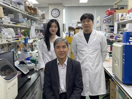
< Photo 1. (From left) Researcher Jungeun Lee, Professor Kwang-Hyun Cho and Postdoctoral Researcher Jeong-Ryeol Gong of the Department of Bio and Brain Engineering at KAIST >
KAIST (represented by President Kwang-Hyung Lee) announced on April 8 that a research team led by Professor Kwang-Hyun Cho from the Department of Bio and Brain Engineering had identified DDX54 as a critical factor that determines the immune evasion capacity of lung cancer cells. They demonstrated that suppressing DDX54 enhances immune cell infiltration into tumors and significantly improves the efficacy of immunotherapy.
Immunotherapy using anti-PD-1 or anti-PD-L1 antibodies is considered a powerful approach in cancer treatment. However, its low response rate limits the number of patients who actually benefit.
To identify likely responders, tumor mutational burden (TMB) has recently been approved by the FDA as a key biomarker for immunotherapy. Cancers with high mutation rates are thought to be more responsive to immune checkpoint inhibitors. However, even tumors with high TMB can display an “immune-desert” phenotype—where immune cell infiltration is severely limited—resulting in poor treatment responses.
< Figure 1. DDX54 was identified as the master regulator that induces resistance to immunotherapy by orchestrating suppression of immune cell infiltration through cancer tissues as lung cancer cells become immune-evasive >
Professor Kwang-Hyun Cho's research team compared transcriptome and genome data of lung cancer patients with immune evasion capabilities through gene regulatory network analysis (A) and discovered DDX54, a master regulator that induces resistance to immunotherapy (B-F).
This study is especially significant in that it successfully demonstrated that suppressing DDX54 in immune-desert lung tumors can overcome immunotherapy resistance and improve treatment outcomes.
The team used transcriptomic and genomic data from immune-evasive lung cancer patients and employed systems biology techniques to infer gene regulatory networks. Through this analysis, they identified DDX54 as a central regulator in the immune evasion of lung cancer cells.
In a syngeneic mouse model, the suppression of DDX54 led to significant increases in the infiltration of anti-cancer immune cells such as T cells and NK cells, and greatly improved the response to immunotherapy.
Single-cell transcriptomic and spatial transcriptomic analyses further showed that combination therapy targeting DDX54 promoted the differentiation of T cells and memory T cells that suppress tumors, while reducing the infiltration of regulatory T cells and exhausted T cells that support tumor growth.
< Figure 2. In the syngeneic mouse model made of lung cancer cells, it was confirmed that inhibiting DDX54 reversed the immune-evasion ability of cancer cells and enhanced the sensitivity to anti-PD-1 therapy >
In a syngeneic mouse model made of lung cancer cells exhibiting immunotherapy resistance, the treatment applied after DDX54 inhibition resulted in statistically significant inhibition of lung cancer growth (B-D) and a significant increase in immune cell infiltration into the tumor tissue (E, F).
The mechanism is believed to involve DDX54 suppression inactivating signaling pathways such as JAK-STAT, MYC, and NF-κB, thereby downregulating immune-evasive proteins CD38 and CD47. This also reduced the infiltration of circulating monocytes—which promote tumor development—and promoted the differentiation of M1 macrophages that play anti-tumor roles.
Professor Kwang-Hyun Cho stated, “We have, for the first time, identified a master regulatory factor that enables immune evasion in lung cancer cells. By targeting this factor, we developed a new therapeutic strategy that can induce responsiveness to immunotherapy in previously resistant cancers.”
He added, “The discovery of DDX54—hidden within the complex molecular networks of cancer cells—was made possible through the systematic integration of systems biology, combining IT and BT.”
The study, led by Professor Kwang-Hyun Cho, was published in the Proceedings of the National Academy of Sciences of the United States of America (PNAS) on April 2, 2025, with Jeong-Ryeol Gong being the first author, Jungeun Lee, a co-first author, and Younghyun Han, a co-author of the article.
< Figure 3. Single-cell transcriptome and spatial transcriptome analysis confirmed that knockdown of DDX54 increased immune cell infiltration into cancer tissues >
In a syngeneic mouse model made of lung cancer cells that underwent immunotherapy in combination with DDX54 inhibition, single-cell transcriptome (H-L) and spatial transcriptome (A-G) analysis of immune cells infiltrating inside cancer tissues were performed. As a result, it was confirmed that anticancer immune cells such as T cells, B cells, and NK cells actively infiltrated the core of lung cancer tissues when DDX54 inhibition and immunotherapy were concurrently administered.
(Paper title: “DDX54 downregulation enhances anti-PD1 therapy in immune-desert lung tumors with high tumor mutational burden,” DOI: https://doi.org/10.1073/pnas.2412310122)
This work was supported by the Ministry of Science and ICT and the National Research Foundation of Korea through the Mid-Career Research Program and Basic Research Laboratory Program.
< Figure 4. The identified master regulator DDX54 was confirmed to induce CD38 and CD47 expression through Jak-Stat3, MYC, and NF-κB activation. >
DDX54 activates the Jak-Stat3, MYC, and NF-κB pathways in lung cancer cells to increase CD38 and CD47 expression (A-G). This creates a cancer microenvironment that contributes to cancer development (H) and ultimately induces immune anticancer treatment resistance.
< Figure 5. It was confirmed that an immune-inflamed environment can be created by combining DDX54 inhibition and immune checkpoint inhibitor (ICI) therapy. >
When DDX54 inhibition and ICI therapy are simultaneously administered, the cancer cell characteristics change, the immune evasion ability is restored, and the environment is transformed into an ‘immune-activated’ environment in which immune cells easily infiltrate cancer tissues. This strengthens the anticancer immune response, thereby increasing the sensitivity of immunotherapy even in lung cancer tissues that previously had low responsiveness to immunotherapy.
2025.04.08 View 20181
-
 KAIST Discovers Molecular Switch that Reverses Cancerous Transformation at the Critical Moment of Transition
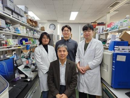
< (From left) PhD student Seoyoon D. Jeong, (bottom) Professor Kwang-Hyun Cho, (top) Dr. Dongkwan Shin, Dr. Jeong-Ryeol Gong >
Professor Kwang-Hyun Cho’s research team has recently been highlighted for their work on developing an original technology for cancer reversal treatment that does not kill cancer cells but only changes their characteristics to reverse them to a state similar to normal cells. This time, they have succeeded in revealing for the first time that a molecular switch that can induce cancer reversal at the moment when normal cells change into cancer cells is hidden in the genetic network.
KAIST (President Kwang-Hyung Lee) announced on the 5th of February that Professor Kwang-Hyun Cho's research team of the Department of Bio and Brain Engineering has succeeded in developing a fundamental technology to capture the critical transition phenomenon at the moment when normal cells change into cancer cells and analyze it to discover a molecular switch that can revert cancer cells back into normal cells.
A critical transition is a phenomenon in which a sudden change in state occurs at a specific point in time, like water changing into steam at 100℃. This critical transition phenomenon also occurs in the process in which normal cells change into cancer cells at a specific point in time due to the accumulation of genetic and epigenetic changes.
The research team discovered that normal cells can enter an unstable critical transition state where normal cells and cancer cells coexist just before they change into cancer cells during tumorigenesis, the production or development of tumors, and analyzed this critical transition state using a systems biology method to develop a cancer reversal molecular switch identification technology that can reverse the cancerization process. They then applied this to colon cancer cells and confirmed through molecular cell experiments that cancer cells can recover the characteristics of normal cells.
This is an original technology that automatically infers a computer model of the genetic network that controls the critical transition of cancer development from single-cell RNA sequencing data, and systematically finds molecular switches for cancer reversion by simulation analysis. It is expected that this technology will be applied to the development of reversion therapies for other cancers in the future.
Professor Kwang-Hyun Cho said, "We have discovered a molecular switch that can revert the fate of cancer cells back to a normal state by capturing the moment of critical transition right before normal cells are changed into an irreversible cancerous state."
< Figure 1. Overall conceptual framework of the technology that automatically constructs a molecular regulatory network from single-cell RNA sequencing data of colon cancer cells to discover molecular switches for cancer reversion through computer simulation analysis. Professor Kwang-Hyun Cho's research team established a fundamental technology for automatic construction of a computer model of a core gene network by analyzing the entire process of tumorigenesis of colon cells turning into cancer cells, and developed an original technology for discovering the molecular switches that can induce cancer cell reversal through attractor landscape analysis. >
He continued, "In particular, this study has revealed in detail, at the genetic network level, what changes occur within cells behind the process of cancer development, which has been considered a mystery until now." He emphasized, "This is the first study to reveal that an important clue that can revert the fate of tumorigenesis is hidden at this very critical moment of change."
< Figure 2. Identification of tumor transition state using single-cell RNA sequencing data from colorectal cancer. Using single-cell RNA sequencing data from colorectal cancer patient-derived organoids for normal and cancerous tissues, a critical transition was identified in which normal and cancerous cells coexist and instability increases (a-d). The critical transition was confirmed to show intermediate levels of major phenotypic features related to cancer or normal tissues that are indicative of the states between the normal and cancerous cells (e). >
The results of this study, conducted by KAIST Dr. Dongkwan Shin (currently at the National Cancer Center), Dr. Jeong-Ryeol Gong, and doctoral student Seoyoon D. Jeong jointly with a research team at Seoul National University that provided the organoids (in vitro cultured tissues) from colon cancer patient, were published as an online paper in the international journal ‘Advanced Science’ published by Wiley on January 22nd.
(Paper title: Attractor landscape analysis reveals a reversion switch in the transition of colorectal tumorigenesis) (DOI: https://doi.org/10.1002/advs.202412503)
< Figure 3. Reconstruction of a dynamic network model for the transition state of colorectal cancer.
A new technology was established to build a gene network computer model that can simulate the dynamic changes between genes by integrating single-cell RNA sequencing data and existing experimental results on gene-to-gene interactions in the critical transition of cancer. (a). Using this technology, a gene network computer model for the critical transition of colorectal cancer was constructed, and the distribution of attractors representing normal and cancer cell phenotypes was investigated through attractor landscape analysis (b-e). >
This study was conducted with the support of the National Research Foundation of Korea under the Ministry of Science and ICT through the Mid-Career Researcher Program and Basic Research Laboratory Program and the Disease-Centered Translational Research Project of the Korea Health Industry Development Institute (KHIDI) of the Ministry of Health and Welfare.
< Figure 4. Quantification of attractor landscapes and discovery of transcription factors for cancer reversibility through perturbation simulation analysis. A methodology for implementing discontinuous attractor landscapes continuously from a computer model of gene networks and quantifying them as cancer scores was introduced (a), and attractor landscapes for the critical transition of colorectal cancer were secured (b-d). By tracking the change patterns of normal and cancer cell attractors through perturbation simulation analysis for each gene, the optimal combination of transcription factors for cancer reversion was discovered (e-h). This was confirmed in various parameter combinations as well (i). >
< Figure 5. Identification and experimental validation of the optimal target gene for cancer reversion. Among the common target genes of the discovered transcription factor combinations, we identified cancer reversing molecular switches that are predicted to suppress cancer cell proliferation and restore the characteristics of normal colon cells (a-d). When inhibitors for the molecular switches were treated to organoids derived from colon cancer patients, it was confirmed that cancer cell proliferation was suppressed and the expression of key genes related to cancer development was inhibited (e-h), and a group of genes related to normal colon epithelium was activated and transformed into a state similar to normal colon cells (i-j). >
< Figure 6. Schematic diagram of the research results. Professor Kwang-Hyun Cho's research team developed an original technology to systematically discover key molecular switches that can induce reversion of colon cancer cells through a systems biology approach using an attractor landscape analysis of a genetic network model for the critical transition at the moment of transformation from normal cells to cancer cells, and verified the reversing effect of actual colon cancer through cellular experiments. >
2025.02.05 View 56545
-
 KAIST Unveils New Possibilities for Treating Intractable Brain Tumors
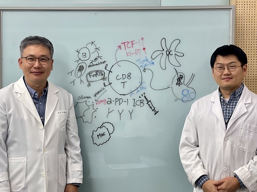
< Photo 1. (From left) Professor Heung Kyu Lee, KAIST Department of Biological Sciences, and Dr. Keun Bon Ku >
Immunotherapy, which enhances the immune system's T cell response to eliminate cancer cells, has emerged as a key approach in cancer treatment. However, in the case of glioblastoma, an aggressive and treatment-resistant brain tumor, numerous clinical trials have failed to confirm their efficacy. Korean researchers have recently analyzed the mechanisms that cause T cell exhaustion, which is characterized by a loss of function or a weakened response following prolonged exposure to antigens in such intractable cancers, identifying key control factors in T cell activation and clarifying the mechanisms that enhance therapeutic effectiveness.
KAIST (represented by President Kwang Hyung Lee) announced on the 6th of November that Professor Heung Kyu Lee’s team from the Department of Biological Sciences, in collaboration with the Korea Research Institute of Chemical Technology (represented by President Young Kuk Lee), has confirmed improved survival rates in a glioblastoma mouse model. By removing the inhibitory Fc gamma receptor (FcγRIIB), the research team was able to restore the responsiveness of cytotoxic T cells to immune checkpoint inhibitors, leading to enhanced anticancer activity.
The research team examined the effect of FcγRIIB, an inhibitory receptor recently found in cytotoxic T cells, on tumor-infiltrating T cells and the therapeutic effectiveness of the anti-PD-1 immune checkpoint inhibitor.
< Figure 1. Study results on improved survival rate due to increased antitumor activity of anti-PD-1 treatment in inhibitory Fc gamma receptor(Fcgr2b) ablation mice with murine glioblastoma. >
Their findings showed that deleting FcγRIIB induced the increase of tumor antigen-specific memory T cells, which helps to suppress exhaustion, enhances stem-like qualities, and reactivates T cell-mediated antitumor immunity, particularly in response to anti-PD-1 treatment. Furthermore, FcγRIIB deletion led to an increase in antigen-specific memory T cells that maintained continuous infiltration into the tumor tissue.
This study presents a new therapeutic target for tumors unresponsive to immune checkpoint inhibitors and demonstrates that combining FcγRIIB inhibition with anti-PD-1 treatment can produce synergistic effects, potentially improving therapeutic outcomes for tumors like glioblastoma, which typically show resistance to anti-PD-1 therapy.
< Figure 2. Overview of the study on the enhanced response to anti-PD-1 therapy for glioblastoma brain tumors upon deletion of the inhibitory Fc gamma receptor (FcγRIIB) in tumor microenvironment. When the inhibitory Fc gamma receptor (FcγRIIB) of cytotoxic T cells is deleted, an increase in tumor-specific memory T cells (Ttsms) was observed. In addition, this T cell subset is identified as originating from the tumor-draining lymph nodes(TdLNs) and leads to persistent infiltration into the tumor tissue. Anti-PD-1 therapy leads to an increased anti-tumor immune response via Ttsms, which is confirmed by increased tumor cell toxicity and increased cell division and decreased cell de-migration indices. Ultimately, the increased cytotoxic T cell immune response leads to an increase in the survival rate of glioblastoma. >
Professor Heung Kyu Lee explained, "This study offers a way to overcome clinical failures in treating brain tumors with immune checkpoint therapy and opens possibilities for broader applications to other intractable cancers. It also highlights the potential of utilizing cytotoxic T cells for tumor cell therapy."
The study, led by Dr. Keun Bon Ku of KAIST (currently a senior researcher at the Korea Research Institute of Chemical Technology's Center for Infectious Disease Diagnosis and Prevention), along with Chae Won Kim, Yumin Kim, Byeong Hoon Kang, Jeongwoo La, In Kang, Won Hyung Park, Stephen Ahn, and Sung Ki Lee, was published online on October 26 in the Journal for ImmunoTherapy of Cancer, an international journal in tumor immunology and therapy from the Society for Immunotherapy of Cancer. (Paper title: “Inhibitory Fcγ receptor deletion enhances CD8 T cell stemness increasing anti-PD-1 therapy responsiveness against glioblastoma,” http://dx.doi.org/10.1136/jitc-2024-009449).
This research received support from the National Research Foundation of Korea, the Bio & Medical Technology Development Program, and the Samsung Science & Technology Foundation.
2024.11.15 View 15538
-
 KAIST team develops smart immune system that can pin down on malignant tumors
A joint research team led by Professor Jung Kyoon Choi of the KAIST Department of Bio and Brain Engineering and Professor Jong-Eun Park of the KAIST Graduate School of Medical Science and Engineering (GSMSE) announced the development of the key technologies to treat cancers using smart immune cells designed based on AI and big data analysis. This technology is expected to be a next-generation immunotherapy that allows precision targeting of tumor cells by having the chimeric antigen receptors (CARs) operate through a logical circuit. Professor Hee Jung An of CHA Bundang Medical Center and Professor Hae-Ock Lee of the Catholic University of Korea also participated in this research to contribute joint effort.
Professor Jung Kyoon Choi’s team built a gene expression database from millions of cells, and used this to successfully develop and verify a deep-learning algorithm that could detect the differences in gene expression patterns between tumor cells and normal cells through a logical circuit. CAR immune cells that were fitted with the logic circuits discovered through this methodology could distinguish between tumorous and normal cells as a computer would, and therefore showed potentials to strike only on tumor cells accurately without causing unwanted side effects.
This research, conducted by co-first authors Dr. Joonha Kwon of the KAIST Department of Bio and Brain Engineering and Ph.D. candidate Junho Kang of KAIST GSMSE, was published by Nature Biotechnology on February 16, under the title Single-cell mapping of combinatorial target antigens for CAR switches using logic gates.
An area in cancer research where the most attempts and advances have been made in recent years is immunotherapy. This field of treatment, which utilizes the patient’s own immune system in order to overcome cancer, has several methods including immune checkpoint inhibitors, cancer vaccines and cellular treatments. Immune cells like CAR-T or CAR-NK equipped with chimera antigen receptors, in particular, can recognize cancer antigens and directly destroy cancer cells.
Starting with its success in blood cancer treatment, scientists have been trying to expand the application of CAR cell therapy to treat solid cancer. But there have been difficulties to develop CAR cells with effective killing abilities against solid cancer cells with minimized side effects. Accordingly, in recent years, the development of smarter CAR engineering technologies, i.e., computational logic gates such as AND, OR, and NOT, to effectively target cancer cells has been underway.
At this point in time, the research team built a large-scale database for cancer and normal cells to discover the exact genes that are expressed only from cancer cells at a single-cell level. The team followed this up by developing an AI algorithm that could search for a combination of genes that best distinguishes cancer cells from normal cells. This algorithm, in particular, has been used to find a logic circuit that can specifically target cancer cells through cell-level simulations of all gene combinations. CAR-T cells equipped with logic circuits discovered through this methodology are expected to distinguish cancerous cells from normal cells like computers, thereby minimizing side effects and maximizing the effects of chemotherapy.
Dr. Joonha Kwon, who is the first author of this paper, said, “this research suggests a new method that hasn’t been tried before. What’s particularly noteworthy is the process in which we found the optimal CAR cell circuit through simulations of millions of individual tumors and normal cells.” He added, “This is an innovative technology that can apply AI and computer logic circuits to immune cell engineering. It would contribute greatly to expanding CAR therapy, which is being successfully used for blood cancer, to solid cancers as well.”
This research was funded by the Original Technology Development Project and Research Program for Next Generation Applied Omic of the Korea Research Foundation.
Figure 1. A schematic diagram of manufacturing and administration process of CAR therapy and of cancer cell-specific dual targeting using CAR.
Figure 2. Deep learning (convolutional neural networks, CNNs) algorithm for selection of dual targets based on gene combination (left) and algorithm for calculating expressing cell fractions by gene combination according to logical circuit (right).
2023.03.09 View 23291
-
 A Genetic Change for Achieving a Long and Healthy Life
Researchers identified a single amino acid change in the tumor suppressor protein in PTEN that extends healthy periods while maintaining longevity
Living a long, healthy life is everyone’s wish, but it is not an easy one to achieve. Many aging studies are developing strategies to increase health spans, the period of life spent with good health, without chronic diseases and disabilities. Researchers at KAIST presented new insights for improving the health span by just regulating the activity of a protein.
A research group under Professor Seung-Jae V. Lee from the Department of Biological Sciences identified a single amino acid change in the tumor suppressor protein phosphatase and tensin homolog (PTEN) that dramatically extends healthy periods while maintaining longevity. This study highlights the importance of the well-conserved tumor suppressor protein PTEN in health span regulation, which can be targeted to develop therapies for promoting healthy longevity in humans. The research was published in Nature Communications on September 24, 2021.
Insulin and insulin-like growth factor-1 (IGF-1) signaling (IIS) is one of the evolutionarily conserved aging-modulatory pathways present in life forms ranging from tiny roundworms to humans. The proper reduction of IIS leads to longevity in animals but often causes defects in multiple health parameters including impaired motility, reproduction, and growth.
The research team found that a specific amino acid change in the PTEN protein improves health status while retaining the longevity conferred by reduced IIS. They used the roundworm C. elegans, an excellent model animal that has been widely used for aging research, mainly because of its very short normal lifespan of about two to three weeks. The PTEN protein is a phosphatase that removes phosphate from lipids as well as proteins. Interestingly, the newly identified amino acid change delicately recalibrated the IIS by partially maintaining protein phosphatase activity while reducing lipid phosphatase activity.
As a result, the amino acid change in the PTEN protein maintained the activity of the longevity-promoting transcription factor Forkhead Box O (FOXO) protein while restricting the detrimental upregulation of another transcription factor, NRF2, leading to long and healthy life in animals with reduced IIS.
Professor Lee said, “Our study raises the exciting possibility of simultaneously promoting longevity and health in humans by slightly tweaking the activity of one protein, PTEN.”
This work was supported by the MInistry of Science and ICT through the National Research Foundation of Korea.
-Publication:Hae-Eun H. Park, Wooseon Hwang, Seokjin Ham, Eunah Kim, Ozlem Altintas, Sangsoon Park, Heehwa G. Son, Yujin Lee, Dongyeop Lee, Won Do Heo, and Seung-Jae V. Lee. 2021. “A PTEN variant uncouples longevity from impaired fitness in Caenorhabditis elegans with reduced insulin/IGF-1 signaling,” Nature Communications, 12(1), 5631. (https://doi.org/10.1038/s41467-021-25920-w)
-ProfileProfessor Seung-Jae V. LeeMolecular Genetics of Aging LaboratoryDepartment of Biological Sciences KAIST
2021.11.19 View 15223
-
 Distinguished Professor Koh Donates His Ho-Am Prize Money
(From left: Distinguished Professor Gou Young Koh and KAIST President Sung-Chul Shin)
Distinguished Professor Gou Young Koh from the Graduate School of Medical Science and Engineering donated one hundred million KRW to KAIST that he received for winning the Ho-Am Prize.
Professor Koh, who is widely renowned for angiogenesis, was appointed as the 2018 laureate of the 28th Ho-Am Prize for demonstrating the effective reduction of tumor progression and metastasis via tumor vessel normalization.
He made the donation to the Graduate School of Medical Science and Engineering, where he conducted his research. “As a basic medical scientist, it is my great honor to receive this prize for the recognition of my research outcome. I will give impetus to research for continuous development,” Professor Koh said.
Professor Koh also received the 5th Asan Award in Medicine in 2012 and the 7th Kyung-Ahm Award in 2011. He was also the awardee of the 17th Wunsch Medical Award. He has donated cash prizes to the school every time he is awarded.
KAIST President Sung-Chul Shin said, “I would like to express my gratitude to the professor for his generous donation to the school. It will be a great help fostering outstanding medical scientists.
Professor Koh received his MD-PhD from the Medical School of Chonbuk National University. After finishing his post-doctoral program at Cornell University and Indiana State University, he was appointed as a professor at Chonbuk National University and POSTECH. Currently, he holds the position of distinguished professor at KAIST and director of the IBS Center for Vascular Research.
2018.06.20 View 9497
-
 Professor Gou Young Koh, 2018 Laureate of Ho-Am Prize
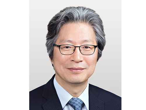
Distinguished Professor Gou Young Koh from the Graduate School of Medical Science and Engineering was appointed a 2018 laureate in medicine of the Ho-Am Prize by the Ho-Am Foundation. Professor Koh is a renowned expert in the field of tumor angiogenesis by exploring the hidden nature of capillary and lymphatic vessels in human organs.
He was recognized for demonstrating the effective reduction of tumor progression and metastasis via tumor vessel normalization. This counterintuitive study result is regarded as a stepping stone for a drug discovery to prevent microvascular diseases.
Besides Professor Koh, Professor Hee Oh from Yale University (Science), Professor Nam-Gyu Park from Sungkyunkwan University (Engineering), Opera Singer Kwangchul Youn (The Arts) and Sister Carla Kang (Community Service) received awards.
The Ho-Am Prize is presented to individuals who have contributed to academics, the arts, and social development, or furthered the welfare of humanity, and commemorates the noble spirit of public service espoused by the late Chairman Byung-chull Lee, who used the pen name Ho-Am.
It was established in 1990 by Kun-Hee Lee, the chairman of Samsung. Awards have been presented to 143 individuals worth a total of 24.4 billion KRW.
2018.04.11 View 15709