%48%65%75%6e%67%20%4b%79%75%20%4c%65%65
-
 Vitamin A Derivative Preserves T Cells’ Function as KAIST Points to a New Strategy for Treating Intractable Brain Tumors
Immune cells can become exhausted after prolonged exposure to cancer, gradually losing their ability to attack tumor cells. This phenomenon is particularly pronounced in aggressive brain tumors and can severely limit the effectiveness of immunotherapy. A KAIST research team has now discovered that all-trans retinoic acid (ATRA), a vitamin A derivative, may help prevent such exhaustion and enhance the efficacy of immune checkpoint inhibitors.
KAIST (President Choongsik Bae) announced on September 8 that a research team led by Professor Heung Kyu Lee from the Department of Biological Sciences, in collaboration with researchers from Seoul St. Mary's Hospital, and Konyang University College of Medicine, has found that all-trans retinoic acid (ATRA), a form of active vitamin A metabolite, suppresses the "terminal exhaustion" of CD8⁺ T cells that attack cancer cells within brain tumors, and can enhance the effect of anti-PD-1 immunotherapy. Anti-PD-1 is a leading immuno-oncology drug (immune checkpoint inhibitor).
Glioblastoma is a representative form of intractable brain cancer that frequently recurs even after surgery, radiation, and chemotherapy. Immune checkpoint inhibitors have shown only limited effect against the disease. One key reason is that immune cells that infiltrate the tumor become exhausted after prolonged combat and lose their functional capacity.
Cells that reach a state of terminal exhaustion in particular lose their ability to kill cancer cells, much like soldiers who have exhausted themselves in prolonged combat. In this state, achieving sufficient therapeutic effect is difficult even with anti-PD-1 therapy, which releases the "brake" cancer cells impose on immune cells.
To address this problem, the research team tried to figure out how to prevent immune cells from reaching total burnout, rather than how to revive the cells. Subsequently, the research team focused on the signaling of ATRA, an active vitamin A metabolite known to regulate cell differentiation and function.
Using an in-vitro model that mimicked the hypoxic, exhaustion-promoting tumor microenvironment of brain tumors, the team induced exhaustion in CD8⁺ T cells. Those conditioned with ATRA progressed to terminal exhaustion at a markedly lower rate. These cells also produced higher levels of immune molecules essential for attacking cancer — including interleukin-2 (IL-2), interferon gamma (IFN-γ), and tumor necrosis factor alpha (TNF-α) — and retained their ability to kill brain tumor cells.
The team also confirmed that ATRA activates WNT/β-catenin signaling within CD8⁺ T cells and increases TCF-1βBD, a TCF-1 isoform important for maintaining T-cell function. In simple terms, the vitamin A metabolite turns on a "function-preserving switch" inside immune cells, helping them avoid complete exhaustion even during prolonged combat with cancer cells.
The effect was also confirmed in mouse glioma models. CD8⁺ T cells conditioned with ATRA maintained better immune function within the tumor, and oral administration of ATRA alone also increased both the number and function of tumor-infiltrating CD8⁺ T cells. As tumor burden decreased, survival was also extended. Notably, in a model of recurrent brain tumors, anti-PD-1 immunotherapy alone showed only limited effect, but combining it with ATRA substantially improved tumor suppression and long-term survival outcomes.
In other words, if anti-PD-1 therapy releases the "brake" imposed on immune cells, ATRA keeps their "battery" from running completely dead. By combining the two approaches, the researchers propose a new combination-therapy strategy that could help overcome the limitations of existing immunotherapy.
The team also analyzed publicly available human glioblastoma datasets. In single-cell transcriptomic data from patients treated with anti-PD-1 therapy, CD8⁺ T cells from responders showed higher retinoic-acid-responsive and WNT signaling gene signatures than those from non-responders. Separately, in an immunotherapy-naïve glioblastoma cohort, patients with a higher proportion of retinoic-acid-responsive CD8⁺ T cells showed significantly better overall survival. The team noted that this study does not directly demonstrate ATRA's therapeutic efficacy in patients, and that further clinical research will be needed to confirm appropriate administration methods, dosages, and combination effects with immunotherapy drugs before it can be applied to actual brain tumor treatment.
Professor Heung Kyu Lee said, "Glioblastoma is one of the cancers most resistant to immunotherapy, because immune cells within the tumor readily become exhausted." He added that the study is meaningful for presenting a molecular mechanism by which active vitamin A signaling helps prevent the terminal exhaustion of CD8⁺ T cells while preserving their anti-cancer function. He also noted that future work validating more precise delivery methods or combination strategies could establish this as a new approach for improving the responsiveness of immunotherapy against intractable brain tumors.
The study was conducted with Dr. In Kang, a postdoctoral researcher in KAIST's Department of Biological Sciences, as first author, and Professor Heung Kyu Lee as corresponding author. Professor Jae-Byum Chang from KAIST's Department of Materials Science and Engineering, Professor Sung Ki Lee from Konyang University College of Medicine, and Professor Stephen Ahn from Seoul St. Mary's Hospital also participated in the research. The findings were published on August 26 in the international journal Signal Transduction and Targeted Therapy.
Paper title: All-trans retinoic acid suppresses CD8⁺ T-cell terminal exhaustion and potentiates anti-PD-1 therapy in glioblastoma
DOI: 10.1038/s41392-026-02852-9
This work was supported by National Research Foundation of Korea grants (RS-2023-NR077244, RS-2024-00439735, RS-2026-25509011 to H.K.L. and RS-2024-00352668 to S.A.). This study was also supported by the Samsung Science and Technology Foundation (SSTF-BA1902-05 to H.K.L.), Republic of Korea. This research was also supported by a grant from the Korea Health Technology R&D Project through the Korea Health Industry Development Institute (KHIDI), funded by the Ministry of Health & Welfare, Republic of Korea (RS-2023-00302794 to J.B.C.).
2026.09.08 View 3974
-
 KAIST Uncovers the “Hidden Key” to Immunotherapy for Intractable Brain Tumors
Researchers have uncovered a clue to why immune checkpoint inhibitors—cancer therapies that release the immune “brakes” exploited by tumors to evade attack—show limited efficacy in some brain tumors. A KAIST research team found that B cell and antibody responses initiated in tumor-draining lymph nodes, rather than T cells alone, are critical to the antitumor effects of anti-CTLA-4 therapy, opening a new avenue for treating intractable brain tumors.
KAIST (President Choongsik Bae) announced on the 19th of July that a research team led by Professor Heung Kyu Lee from the Department of Biological Sciences has identified a previously unrecognized immune mechanism through which anti-CTLA-4, a type of immune checkpoint inhibitor, promotes B-cell responses in tumor-draining lymph nodes, thereby helping the immune system attack brain tumors.
Glioblastoma is one of the most aggressive malignant brain tumors, with frequent recurrence and a poor prognosis even after surgery and radiation therapy. Immune checkpoint inhibitors, which restore the ability of immune cells to attack cancer cells, have produced substantial therapeutic benefits in various cancers. However, their effectiveness in glioblastoma has remained limited because of the highly immunosuppressive environment surrounding the tumor.
Researchers have traditionally regarded T cells—immune cells that can directly attack cancer cells—as the primary target of immune checkpoint inhibitors. B cells, meanwhile, are well known for producing antibodies following infection or vaccination, but their role in brain tumor immunotherapy has remained largely unexplored. The research team therefore investigated whether anti-CTLA-4 could influence B-cell responses as well as T-cell responses.
The findings challenged the prevailing T-cell-centered view. In mouse glioma models, anti-CTLA-4 treatment reduced tumor burden and significantly prolonged survival. However, these therapeutic effects were largely lost in mice lacking B cells, demonstrating that B cells are required for the efficacy of anti-CTLA-4 treatment in these models.
The team also identified where B cells played their key role. Rather than being prominent in the brain, where the tumor cells were located, the response increased markedly in the Deep Cervical Lymph Nodes, which are located deep in the neck and receive lymphatic drainage from the brain. In particular, germinal center B cells and T follicular helper cells, both of which are important for antibody formation, increased together in these lymph nodes. This was accompanied by an increase in immunoglobulin G, or IgG, responses. IgG is a major class of antibody that can recognize cancer cells as targets and help immune cells eliminate them.
The resulting IgG antibodies bound to the surface of glioma cells, helping macrophages—immune cells that engulf foreign substances and cancer cells—remove the tumor cells more effectively.
The research team also examined this process directly in vivo. Using a specialized dual-reporter glioma model expressing the red fluorescent protein mCherry and the green fluorescent protein EGFP, the researchers successfully visualized tumor-infiltrating phagocytes actively engulfing glioma cells following anti-CTLA-4 treatment.
The study provides the first functional evidence that B-cell immune responses—previously known mainly for their roles in infection and vaccination—can be a key factor determining the effectiveness of immunotherapy for hard-to-treat brain tumors. It also expands the conventional T-cell-centered framework of cancer immunotherapy by showing that treatment efficacy can be strongly shaped by immune responses originating not only within the tumor, but also in tumor-draining lymph nodes outside it.
Yumin Kim, a postdoctoral researcher in the KAIST Department of Biological Sciences, served as the first author of the study, with Professor Heung Kyu Lee serving as the corresponding author. Professor Ji Eun Oh from the KAIST Graduate School of Medical Science and Engineering also contributed to the research. The findings were published on July 10 in Science Immunology.
Paper title: The efficacy of immunotherapy in glioma requires distal B-cell responses in tumor-draining lymph nodes
DOI: 10.1126/sciimmunol.adz2494
Authors: Yumin Kim, In Kang, Byeong Hoon Kang, Won Hyung Park, Chae Won Kim, Hyun-Jin Kim, Jeongwoo La, Myoung Seung Kwon, Sang Hee Park, Seo Hyeon Im, Hyeon Cheol Kim, Keun Bon Ku, Minji Kim, and Ji Eun Oh from KAIST, with Heung Kyu Lee from KAIST as the corresponding author.
This research was supported by National Research Foundation of Korea grants RS-2023-NR077244 (to H.K.L.), RS-2024-00439735 (to H.K.L.), RS-2024-00411928 (to Y.K.), RS-2025-00517107 (to J.E.O.), and RS-2026-25509011 (to H.K.L.). This study was also supported by Samsung Science and Technology Foundation grants SSTF-BA1902-05 (to H.K.L.) and SSTF-BA2201-11 (to J.E.O.).
2026.07.20 View 5549
-
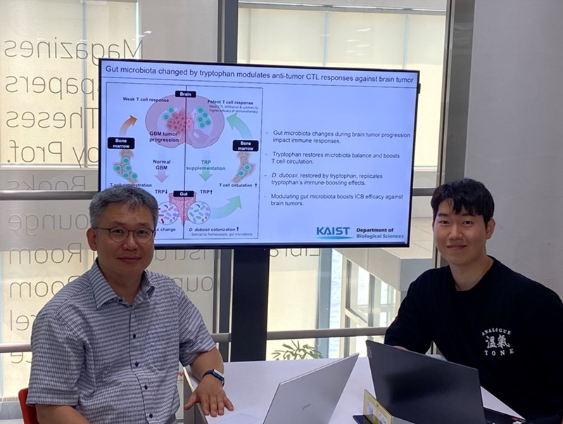 KAIST Enhances Immunotherapy for Difficult-to-Treat Brain Tumors with Gut Microbiota
< Photo 1.(From left) Prof. Heung Kyu Lee, Department of Biological Sciences,
and Dr. Hyeon Cheol Kim>
Advanced treatments, known as immunotherapies that activate T cells—our body's immune cells—to eliminate cancer cells, have shown limited efficacy as standalone therapies for glioblastoma, the most lethal form of brain tumor. This is due to their minimal response to glioblastoma and high resistance to treatment.
Now, a KAIST research team has now demonstrated a new therapeutic strategy that can enhance the efficacy of immunotherapy for brain tumors by utilizing gut microbes and their metabolites. This also opens up possibilities for developing microbiome-based immunotherapy supplements in the future.
KAIST (President Kwang Hyung Lee) announced on July 1 that a research team led by Professor Heung Kyu Lee of the Department of Biological Sciences discovered and demonstrated a method to significantly improve the efficiency of glioblastoma immunotherapy by focusing on changes in the gut microbial ecosystem.
The research team noted that as glioblastoma progresses, the concentration of ‘tryptophan’, an important amino acid in the gut, sharply decreases, leading to changes in the gut microbial ecosystem. They discovered that by supplementing tryptophan to restore microbial diversity, specific beneficial strains activate CD8 T cells (a type of immune cell) and induce their infiltration into tumor tissues. Through a mouse model of glioblastoma, the research team confirmed that tryptophan supplementation enhanced the response of cancer-attacking T cells (especially CD8 T cells), leading to their increased migration to tumor sites such as lymph nodes and the brain.
In this process, they also revealed that ‘Duncaniella dubosii’, a beneficial commensal bacterium present in the gut, plays a crucial role. This bacterium helped T cells effectively redistribute within the body, and survival rates significantly improved when used in combination with immunotherapy (anti-PD-1).
Furthermore, it was demonstrated that even when this commensal bacterium was administered alone to germ-free mice (mice without any commensal microbes), the survival rate for glioblastoma increased. This is because the bacterium utilizes tryptophan to regulate the gut environment, and the metabolites produced in this process strengthen the ability of CD8 T cells to attack cancer cells.
Professor Heung Kyu Lee explained, "This research is a meaningful achievement, showing that even in intractable brain tumors where immune checkpoint inhibitors had no effect, a combined strategy utilizing gut microbes can significantly enhance treatment response."
Dr. Hyeon Cheol Kim of KAIST (currently a postdoctoral researcher at the Institute for Biological Sciences) participated as the first author. The research findings were published online in Cell Reports, an international journal in the life sciences, on June 26.
This research was conducted as part of the Basic Research Program and Bio & Medical Technology Development Program supported by the Ministry of Science and ICT and the National Research Foundation of Korea.
※Paper Title: Gut microbiota dysbiosis induced by brain tumor modulates the efficacy of immunotherapy
※DOI: https://doi.org/10.1016/j.celrep.2025.115825
2025.07.02 View 11276
-
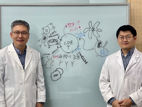 KAIST Unveils New Possibilities for Treating Intractable Brain Tumors
< Photo 1. (From left) Professor Heung Kyu Lee, KAIST Department of Biological Sciences, and Dr. Keun Bon Ku >
Immunotherapy, which enhances the immune system's T cell response to eliminate cancer cells, has emerged as a key approach in cancer treatment. However, in the case of glioblastoma, an aggressive and treatment-resistant brain tumor, numerous clinical trials have failed to confirm their efficacy. Korean researchers have recently analyzed the mechanisms that cause T cell exhaustion, which is characterized by a loss of function or a weakened response following prolonged exposure to antigens in such intractable cancers, identifying key control factors in T cell activation and clarifying the mechanisms that enhance therapeutic effectiveness.
KAIST (represented by President Kwang Hyung Lee) announced on the 6th of November that Professor Heung Kyu Lee’s team from the Department of Biological Sciences, in collaboration with the Korea Research Institute of Chemical Technology (represented by President Young Kuk Lee), has confirmed improved survival rates in a glioblastoma mouse model. By removing the inhibitory Fc gamma receptor (FcγRIIB), the research team was able to restore the responsiveness of cytotoxic T cells to immune checkpoint inhibitors, leading to enhanced anticancer activity.
The research team examined the effect of FcγRIIB, an inhibitory receptor recently found in cytotoxic T cells, on tumor-infiltrating T cells and the therapeutic effectiveness of the anti-PD-1 immune checkpoint inhibitor.
< Figure 1. Study results on improved survival rate due to increased antitumor activity of anti-PD-1 treatment in inhibitory Fc gamma receptor(Fcgr2b) ablation mice with murine glioblastoma. >
Their findings showed that deleting FcγRIIB induced the increase of tumor antigen-specific memory T cells, which helps to suppress exhaustion, enhances stem-like qualities, and reactivates T cell-mediated antitumor immunity, particularly in response to anti-PD-1 treatment. Furthermore, FcγRIIB deletion led to an increase in antigen-specific memory T cells that maintained continuous infiltration into the tumor tissue.
This study presents a new therapeutic target for tumors unresponsive to immune checkpoint inhibitors and demonstrates that combining FcγRIIB inhibition with anti-PD-1 treatment can produce synergistic effects, potentially improving therapeutic outcomes for tumors like glioblastoma, which typically show resistance to anti-PD-1 therapy.
< Figure 2. Overview of the study on the enhanced response to anti-PD-1 therapy for glioblastoma brain tumors upon deletion of the inhibitory Fc gamma receptor (FcγRIIB) in tumor microenvironment. When the inhibitory Fc gamma receptor (FcγRIIB) of cytotoxic T cells is deleted, an increase in tumor-specific memory T cells (Ttsms) was observed. In addition, this T cell subset is identified as originating from the tumor-draining lymph nodes(TdLNs) and leads to persistent infiltration into the tumor tissue. Anti-PD-1 therapy leads to an increased anti-tumor immune response via Ttsms, which is confirmed by increased tumor cell toxicity and increased cell division and decreased cell de-migration indices. Ultimately, the increased cytotoxic T cell immune response leads to an increase in the survival rate of glioblastoma. >
Professor Heung Kyu Lee explained, "This study offers a way to overcome clinical failures in treating brain tumors with immune checkpoint therapy and opens possibilities for broader applications to other intractable cancers. It also highlights the potential of utilizing cytotoxic T cells for tumor cell therapy."
The study, led by Dr. Keun Bon Ku of KAIST (currently a senior researcher at the Korea Research Institute of Chemical Technology's Center for Infectious Disease Diagnosis and Prevention), along with Chae Won Kim, Yumin Kim, Byeong Hoon Kang, Jeongwoo La, In Kang, Won Hyung Park, Stephen Ahn, and Sung Ki Lee, was published online on October 26 in the Journal for ImmunoTherapy of Cancer, an international journal in tumor immunology and therapy from the Society for Immunotherapy of Cancer. (Paper title: “Inhibitory Fcγ receptor deletion enhances CD8 T cell stemness increasing anti-PD-1 therapy responsiveness against glioblastoma,” http://dx.doi.org/10.1136/jitc-2024-009449).
This research received support from the National Research Foundation of Korea, the Bio & Medical Technology Development Program, and the Samsung Science & Technology Foundation.
2024.11.15 View 15870
-
 Biomarker Predicts Who Will Have Severe COVID-19
- Airway cell analyses showing an activated immune axis could pinpoint the COVID-19 patients who will most benefit from targeted therapies.-
KAIST researchers have identified key markers that could help pinpoint patients who are bound to get a severe reaction to COVID-19 infection. This would help doctors provide the right treatments at the right time, potentially saving lives. The findings were published in the journal Frontiers in Immunology on August 28.
People’s immune systems react differently to infection with SARS-CoV-2, the virus that causes COVID-19, ranging from mild to severe, life-threatening responses.
To understand the differences in responses, Professor Heung Kyu Lee and PhD candidate Jang Hyun Park from the Graduate School of Medical Science and Engineering at KAIST analysed ribonucleic acid (RNA) sequencing data extracted from individual airway cells of healthy controls and of mildly and severely ill patients with COVID-19. The data was available in a public database previously published by a group of Chinese researchers.
“Our analyses identified an association between immune cells called neutrophils and special cell receptors that bind to the steroid hormone glucocorticoid,” Professor Lee explained. “This finding could be used as a biomarker for predicting disease severity in patients and thus selecting a targeted therapy that can help treat them at an appropriate time,” he added.
Severe illness in COVID-19 is associated with an exaggerated immune response that leads to excessive airway-damaging inflammation. This condition, known as acute respiratory distress syndrome (ARDS), accounts for 70% of deaths in fatal COVID-19 infections.
Scientists already know that this excessive inflammation involves heightened neutrophil recruitment to the airways, but the detailed mechanisms of this reaction are still unclear.
Lee and Park’s analyses found that a group of immune cells called myeloid cells produced excess amounts of neutrophil-recruiting chemicals in severely ill patients, including a cytokine called tumour necrosis factor (TNF) and a chemokine called CXCL8.
Further RNA analyses of neutrophils in severely ill patients showed they were less able to recruit very important T cells needed for attacking the virus. At the same time, the neutrophils produced too many extracellular molecules that normally trap pathogens, but damage airway cells when produced in excess.
The researchers additionally found that the airway cells in severely ill patients were not expressing enough glucocorticoid receptors. This was correlated with increased CXCL8 expression and neutrophil recruitment.
Glucocorticoids, like the well-known drug dexamethasone, are anti-inflammatory agents that could play a role in treating COVID-19. However, using them in early or mild forms of the infection could suppress the necessary immune reactions to combat the virus. But if airway damage has already happened in more severe cases, glucocorticoid treatment would be ineffective.
Knowing who to give this treatment to and when is really important. COVID-19 patients showing reduced glucocorticoid receptor expression, increased CXCL8 expression, and excess neutrophil recruitment to the airways could benefit from treatment with glucocorticoids to prevent airway damage. Further research is needed, however, to confirm the relationship between glucocorticoids and neutrophil inflammation at the protein level.
“Our study could serve as a springboard towards more accurate and reliable COVID-19 treatments,” Professor Lee said.
This work was supported by the National Research Foundation of Korea, and Mobile Clinic Module Project funded by KAIST.
Figure. Low glucocorticoid receptor (GR) expression led to excessive inflammation and lung damage by neutrophils through enhancing the expression of CXCL8 and other cytokines.
Image credit: Professor Heung Kyu Lee, KAIST. Created with Biorender.com.
Image usage restrictions: News organizations may use or redistribute these figures and image, with proper attribution, as part of news coverage of this paper only.
-Publication:
Jang Hyun Park, and Heung Kyu Lee. (2020). Re-analysis of Single Cell Transcriptome Reveals That the NR3C1-CXCL8-Neutrophil Axis Determines the Severity of COVID-19. Frontiers in Immunology, Available online at https://doi.org/10.3389/fimmu.2020.02145
-Profile: Heung Kyu Lee
Associate Professor
heungkyu.lee@kaist.ac.kr
https://www.heungkyulee.kaist.ac.kr/
Laboratory of Host Defenses
Graduate School of Medical Science and Engineering (GSMSE)
The Center for Epidemic Preparedness at KAIST Institute
http://kaist.ac.kr
Korea Advanced Institute of Science and Technology (KAIST)
Daejeon, Republic of Korea
Profile: Jang Hyun Park
PhD Candidate
janghyun.park@kaist.ac.kr
GSMSE, KAIST
2020.09.17 View 30118