%4a%69%6e%2d%48%65%65%20%48%61%6e
-
 Why Do Dementia and Cognitive Decline Patients Remain Stuck in Past Memories?… KAIST Identifies Memory-Switching Mechanism
<(From Left) Professor Jin-Hee Han, Dr. Mujun Kim>
“Why do patients with dementia or cognitive decline remain stuck in past memories?”
KAIST researchers have identified, for the first time in the world, the existence of a “neural switch” in the brain that selectively retrieves the most recent memories. This study reveals the principle by which the brain selects necessary information between past memories and new memories, presenting new possibilities for future treatments for memory decline and reduced cognitive flexibility.
KAIST announced on the 17th of May that a research team led by Professor Jin-Hee Han of the Department of Biological Sciences has discovered, for the first time in the world, that a specific neural circuit connecting the medial septum (MS, a brain region that regulates memory and learning) and the medial entorhinal cortex (MEC, a brain region connected to the hippocampus* that processes memory information) switches between past and recent memories and plays a key role in selecting up-to-date information appropriate for the situation.
*Hippocampus: a key brain region that creates and stores new memories
We live by updating our memories through new experiences every day. For example, if the restaurant we visited today was more satisfying than the one we visited yesterday, the brain modifies the existing memory to reflect the new information. In this way, the ability to select necessary information between past and present memories is central to higher cognitive functions such as decision-making, problem-solving, future prediction, and logical reasoning. However, the principle by which the brain distinguishes and switches between memories has long remained unknown.
The research team focused on the medial septum, located deep within the brain. The medial septum regulates the activity rhythms of the hippocampus and acts as a “conductor” that helps the brain effectively store and retrieve information.
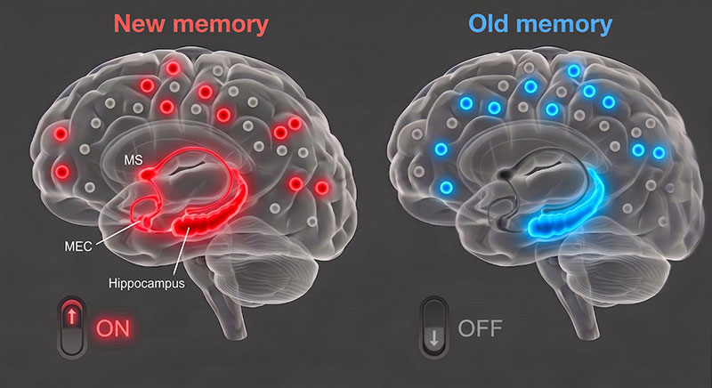
The study found that when specific neurons in the medial septum send signals to the medial entorhinal cortex, a brain region that processes memory information and delivers it to the hippocampus, the brain is better able to recall recent memories.
<(AI image) An inhibitory neural circuit switch in the MS–MEC pathway that regulates the selection between past and recent memories>
Conversely, when the research team artificially blocked this neural circuit using light, experimental animals were unable to use recent information and behaved according to past patterns. Neural activity in the hippocampus, which plays an important role in memory representation, also reverted to a past state. This shows that the circuit acts as a “neural switch” that selects the most recent information needed for the current situation among multiple memories.
The research team also analyzed memory performance according to brain activity states. Our brain repeatedly shifts between an “online state” (theta waves, brain waves activated during learning and concentration), in which it actively processes information, and an “offline state” (delta waves, slow brain waves that appear during sleep or rest), which is a resting state.
The analysis showed that the longer the online state was maintained, the better recent memories were recalled, while frequent switching between online and offline states significantly reduced memory retrieval ability. This suggests that specific brain rhythms and states are important neurobiological indicators that determine effective memory retrieval.
This study is significant in that it identified the mechanism by which the brain flexibly reflects new information while maintaining past memories. The research team expects that this discovery could lead to the development of new therapeutic technologies to improve memory decline and reduced cognitive flexibility in patients with degenerative brain diseases such as dementia and Alzheimer’s disease.
<An inhibitory neural circuit switch in the MS–MEC pathway that regulates the selection between past and recent memories>
Professor Jin-Hee Han stated, “This study presents a new paradigm for understanding the principle by which our brain organizes and uses numerous experiences in chronological order,” adding, “Previously, memory retrieval was understood simply as the replaying of stored traces, but through this study, we proved that the brain has a regulatory system that actively selects recent information among competing memories.”
This study involved Dr. Mujun Kim of the Department of Biological Sciences at KAIST, doctoral students Boin Suh, Sunhoi So, Jung Wook Choi, Jaemin Hwang, and Juhee Park, and was published on April 29 in Nature Neuroscience, a top-tier international journal in neuroscience.
※ Paper title: “A septo-entorhinal GABAergic pathway that enables switching between episodic memories,” https://doi.org/10.1038/s41593-026-02280-6 ※ Author information: Mujun Kim (KAIST, first author), Boin Suh (KAIST), Sunhoi So (KAIST), Jung Wook Choi (KAIST), Jaemin Hwang (KAIST), Juhee Park (KAIST) & Jin-Hee Han (KAIST, corresponding author)
This research was supported by the Mid-Career Research Program (National Research Foundation of Korea), the Samsung Science and Technology Foundation, and the KAIST Jang Young Sil Fellow Program.
2026.05.18 View 2517
-
 Decoding Fear: KAIST Identifies An Affective Brain Circuit Crucial for Fear Memory Formation by Non-nociceptive Threat Stimulus
Fear memories can form in the brain following exposure to threatening situations such as natural disasters, accidents, or violence. When these memories become excessive or distorted, they can lead to severe mental health disorders, including post-traumatic stress disorder (PTSD), anxiety disorders, and depression. However, the mechanisms underlying fear memory formation triggered by affective pain rather than direct physical pain have remained largely unexplored – until now.
A KAIST research team has identified, for the first time, a brain circuit specifically responsible for forming fear memories in the absence of physical pain, marking a significant advance in understanding how psychological distress is processed and drives fear memory formation in the brain. This discovery opens the door to the development of targeted treatments for trauma-related conditions by addressing the underlying neural pathways.
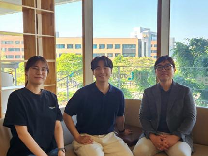
< Photo 1. (from left) Professor Jin-Hee Han, Dr. Junho Han and Ph.D. Candidate Boin Suh of the Department of Biological Sciences >
KAIST (President Kwang-Hyung Lee) announced on May 15th that the research team led by Professor Jin-Hee Han in the Department of Biological Sciences has identified the pIC-PBN circuit*, a key neural pathway involved in forming fear memories triggered by psychological threats in the absence of sensory pain. This groundbreaking work was conducted through experiments with mice.*pIC–PBN circuit: A newly identified descending neural pathway from the posterior insular cortex (pIC) to the parabrachial nucleus (PBN), specialized for transmitting psychological threat information.
Traditionally, the lateral parabrachial nucleus (PBN) has been recognized as a critical part of the ascending pain pathway, receiving pain signals from the spinal cord. However, this study reveals a previously unknown role for the PBN in processing fear induced by non-painful psychological stimuli, fundamentally changing our understanding of its function in the brain.
This work is considered the first experimental evidence that 'emotional distress' and 'physical pain' are processed through different neural circuits to form fear memories, making it a significant contribution to the field of neuroscience. It clearly demonstrates the existence of a dedicated pathway (pIC-PBN) for transmitting emotional distress.
The study's first author, Dr. Junho Han, shared the personal motivation behind this research: “Our dog, Lego, is afraid of motorcycles. He never actually crashed into one, but ever since having a traumatizing event of having a motorbike almost run into him, just hearing the sound now triggers a fearful response. Humans react similarly – even if you didn’t have a personal experience of being involved in an accident, a near-miss or exposure to alarming media can create lasting fear memories, which may eventually lead to PTSD.”
He continued, “Until now, fear memory research has mainly relied on experimental models involving physical pain. However, much of real-world human fears arise from psychological threats, rather than from direct physical harm. Despite this, little was known about the brain circuits responsible for processing these psychological threats that can drive fear memory formation.”
To investigate this, the research team developed a novel fear conditioning model that utilizes visual threat stimuli instead of electrical shocks. In this model, mice were exposed to a rapidly expanding visual disk on a ceiling screen, simulating the threat of an approaching predator. This approach allowed the team to demonstrate that fear memories can form in response to a non-nociceptive, psychological threat alone, without the need for physical pain.
< Figure 1. Artificial activation of the posterior insular cortex (pIC) to lateral parabrachial nucleus (PBN) neural circuit induces anxiety-like behaviors and fear memory formation in mice. >
Using advanced chemogenetic and optogenetic techniques, the team precisely controlled neuronal activity, revealing that the lateral parabrachial nucleus (PBN) is essential to form fear memories in response to visual threats. They further traced the origin of these signals to the posterior insular cortex (pIC), a region known to process negative emotions and pain, confirming a direct connection between the two areas.
The study also showed that inhibiting the pIC–PBN circuit significantly reduced fear memory formation in response to visual threats, without affecting innate fear responses or physical pain-based learning. Conversely, artificially activating this circuit alone was sufficient to drive fear memory formation, confirming its role as a key pathway for processing psychological threat information.
< Figure 2. Schematic diagram of brain neural circuits transmitting emotional & physical pain threat signals. Visual threat stimuli do not involve physical pain but can create an anxious state and form fear memory through the affective pain signaling pathway. >
Professor Jin-Hee Han commented, “This study lays an important foundation for understanding how emotional distress-based mental disorders, such as PTSD, panic disorder, and anxiety disorder, develop, and opens new possibilities for targeted treatment approaches.”
The findings, authored by Dr. Junho Han (first author), Ph.D. candidate Boin Suh (second author), and Dr. Jin-Hee Han (corresponding author) of the Department of Biological Sciences, were published online in the international journal Science Advances on May 9, 2025.※ Paper Title: A top-down insular cortex circuit crucial for non-nociceptive fear learning. Science Advances (https://doi.org/10.1101/2024.10.14.618356)※ Author Information: Junho Han (first author), Boin Suh (second author), and Jin-Hee Han (corresponding author)
This research was supported by grants from the National Research Foundation of Korea (NRF-2022M3E5E8081183 and NRF-2017M3C7A1031322).
2025.05.15 View 14176