%44%65%70%61%72%74%6d%65%6e%74%20%6f%66%20%42%69%6f%6c%6f%67%69%63%61%6c%20%53%63%69%65%6e%63%65
-
 Why Do Dementia and Cognitive Decline Patients Remain Stuck in Past Memories?… KAIST Identifies Memory-Switching Mechanism
<(From Left) Professor Jin-Hee Han, Dr. Mujun Kim>
“Why do patients with dementia or cognitive decline remain stuck in past memories?”
KAIST researchers have identified, for the first time in the world, the existence of a “neural switch” in the brain that selectively retrieves the most recent memories. This study reveals the principle by which the brain selects necessary information between past memories and new memories, presenting new possibilities for future treatments for memory decline and reduced cognitive flexibility.
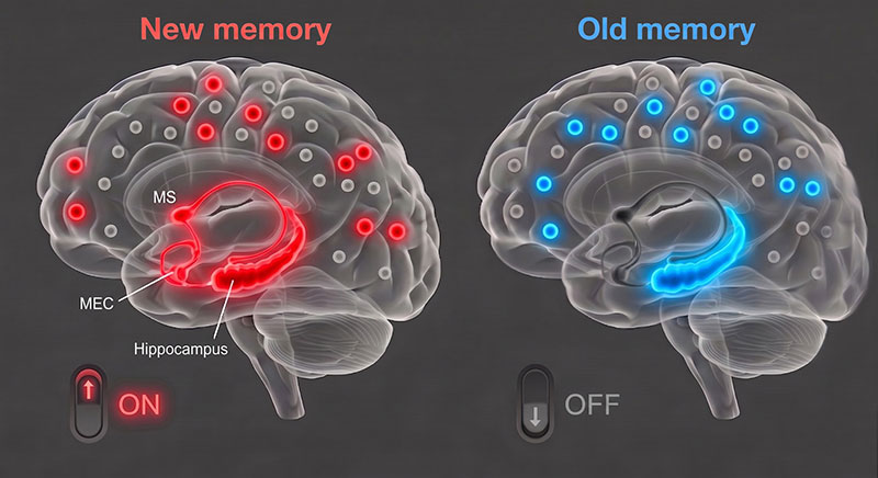
KAIST announced on the 17th of May that a research team led by Professor Jin-Hee Han of the Department of Biological Sciences has discovered, for the first time in the world, that a specific neural circuit connecting the medial septum (MS, a brain region that regulates memory and learning) and the medial entorhinal cortex (MEC, a brain region connected to the hippocampus* that processes memory information) switches between past and recent memories and plays a key role in selecting up-to-date information appropriate for the situation.
*Hippocampus: a key brain region that creates and stores new memories
We live by updating our memories through new experiences every day. For example, if the restaurant we visited today was more satisfying than the one we visited yesterday, the brain modifies the existing memory to reflect the new information. In this way, the ability to select necessary information between past and present memories is central to higher cognitive functions such as decision-making, problem-solving, future prediction, and logical reasoning. However, the principle by which the brain distinguishes and switches between memories has long remained unknown.
The research team focused on the medial septum, located deep within the brain. The medial septum regulates the activity rhythms of the hippocampus and acts as a “conductor” that helps the brain effectively store and retrieve information.
The study found that when specific neurons in the medial septum send signals to the medial entorhinal cortex, a brain region that processes memory information and delivers it to the hippocampus, the brain is better able to recall recent memories.
<(AI image) An inhibitory neural circuit switch in the MS–MEC pathway that regulates the selection between past and recent memories>
Conversely, when the research team artificially blocked this neural circuit using light, experimental animals were unable to use recent information and behaved according to past patterns. Neural activity in the hippocampus, which plays an important role in memory representation, also reverted to a past state. This shows that the circuit acts as a “neural switch” that selects the most recent information needed for the current situation among multiple memories.
The research team also analyzed memory performance according to brain activity states. Our brain repeatedly shifts between an “online state” (theta waves, brain waves activated during learning and concentration), in which it actively processes information, and an “offline state” (delta waves, slow brain waves that appear during sleep or rest), which is a resting state.
The analysis showed that the longer the online state was maintained, the better recent memories were recalled, while frequent switching between online and offline states significantly reduced memory retrieval ability. This suggests that specific brain rhythms and states are important neurobiological indicators that determine effective memory retrieval.
This study is significant in that it identified the mechanism by which the brain flexibly reflects new information while maintaining past memories. The research team expects that this discovery could lead to the development of new therapeutic technologies to improve memory decline and reduced cognitive flexibility in patients with degenerative brain diseases such as dementia and Alzheimer’s disease.
<An inhibitory neural circuit switch in the MS–MEC pathway that regulates the selection between past and recent memories>
Professor Jin-Hee Han stated, “This study presents a new paradigm for understanding the principle by which our brain organizes and uses numerous experiences in chronological order,” adding, “Previously, memory retrieval was understood simply as the replaying of stored traces, but through this study, we proved that the brain has a regulatory system that actively selects recent information among competing memories.”
This study involved Dr. Mujun Kim of the Department of Biological Sciences at KAIST, doctoral students Boin Suh, Sunhoi So, Jung Wook Choi, Jaemin Hwang, and Juhee Park, and was published on April 29 in Nature Neuroscience, a top-tier international journal in neuroscience.
※ Paper title: “A septo-entorhinal GABAergic pathway that enables switching between episodic memories,” https://doi.org/10.1038/s41593-026-02280-6 ※ Author information: Mujun Kim (KAIST, first author), Boin Suh (KAIST), Sunhoi So (KAIST), Jung Wook Choi (KAIST), Jaemin Hwang (KAIST), Juhee Park (KAIST) & Jin-Hee Han (KAIST, corresponding author)
This research was supported by the Mid-Career Research Program (National Research Foundation of Korea), the Samsung Science and Technology Foundation, and the KAIST Jang Young Sil Fellow Program.
2026.05.18 View 215
Why Do Dementia and Cognitive Decline Patients Remain Stuck in Past Memories?… KAIST Identifies Memory-Switching Mechanism
<(From Left) Professor Jin-Hee Han, Dr. Mujun Kim>
“Why do patients with dementia or cognitive decline remain stuck in past memories?”
KAIST researchers have identified, for the first time in the world, the existence of a “neural switch” in the brain that selectively retrieves the most recent memories. This study reveals the principle by which the brain selects necessary information between past memories and new memories, presenting new possibilities for future treatments for memory decline and reduced cognitive flexibility.
KAIST announced on the 17th of May that a research team led by Professor Jin-Hee Han of the Department of Biological Sciences has discovered, for the first time in the world, that a specific neural circuit connecting the medial septum (MS, a brain region that regulates memory and learning) and the medial entorhinal cortex (MEC, a brain region connected to the hippocampus* that processes memory information) switches between past and recent memories and plays a key role in selecting up-to-date information appropriate for the situation.
*Hippocampus: a key brain region that creates and stores new memories
We live by updating our memories through new experiences every day. For example, if the restaurant we visited today was more satisfying than the one we visited yesterday, the brain modifies the existing memory to reflect the new information. In this way, the ability to select necessary information between past and present memories is central to higher cognitive functions such as decision-making, problem-solving, future prediction, and logical reasoning. However, the principle by which the brain distinguishes and switches between memories has long remained unknown.
The research team focused on the medial septum, located deep within the brain. The medial septum regulates the activity rhythms of the hippocampus and acts as a “conductor” that helps the brain effectively store and retrieve information.
The study found that when specific neurons in the medial septum send signals to the medial entorhinal cortex, a brain region that processes memory information and delivers it to the hippocampus, the brain is better able to recall recent memories.
<(AI image) An inhibitory neural circuit switch in the MS–MEC pathway that regulates the selection between past and recent memories>
Conversely, when the research team artificially blocked this neural circuit using light, experimental animals were unable to use recent information and behaved according to past patterns. Neural activity in the hippocampus, which plays an important role in memory representation, also reverted to a past state. This shows that the circuit acts as a “neural switch” that selects the most recent information needed for the current situation among multiple memories.
The research team also analyzed memory performance according to brain activity states. Our brain repeatedly shifts between an “online state” (theta waves, brain waves activated during learning and concentration), in which it actively processes information, and an “offline state” (delta waves, slow brain waves that appear during sleep or rest), which is a resting state.
The analysis showed that the longer the online state was maintained, the better recent memories were recalled, while frequent switching between online and offline states significantly reduced memory retrieval ability. This suggests that specific brain rhythms and states are important neurobiological indicators that determine effective memory retrieval.
This study is significant in that it identified the mechanism by which the brain flexibly reflects new information while maintaining past memories. The research team expects that this discovery could lead to the development of new therapeutic technologies to improve memory decline and reduced cognitive flexibility in patients with degenerative brain diseases such as dementia and Alzheimer’s disease.
<An inhibitory neural circuit switch in the MS–MEC pathway that regulates the selection between past and recent memories>
Professor Jin-Hee Han stated, “This study presents a new paradigm for understanding the principle by which our brain organizes and uses numerous experiences in chronological order,” adding, “Previously, memory retrieval was understood simply as the replaying of stored traces, but through this study, we proved that the brain has a regulatory system that actively selects recent information among competing memories.”
This study involved Dr. Mujun Kim of the Department of Biological Sciences at KAIST, doctoral students Boin Suh, Sunhoi So, Jung Wook Choi, Jaemin Hwang, and Juhee Park, and was published on April 29 in Nature Neuroscience, a top-tier international journal in neuroscience.
※ Paper title: “A septo-entorhinal GABAergic pathway that enables switching between episodic memories,” https://doi.org/10.1038/s41593-026-02280-6 ※ Author information: Mujun Kim (KAIST, first author), Boin Suh (KAIST), Sunhoi So (KAIST), Jung Wook Choi (KAIST), Jaemin Hwang (KAIST), Juhee Park (KAIST) & Jin-Hee Han (KAIST, corresponding author)
This research was supported by the Mid-Career Research Program (National Research Foundation of Korea), the Samsung Science and Technology Foundation, and the KAIST Jang Young Sil Fellow Program.
2026.05.18 View 215 -
 3D Stem Cell Culture Technology to Shift the Paradigm of Regenerative Medicine
< (From left) KAIST Dr. Changjin Seo, Professor Sangyong Jon >
A breakthrough technology has been developed to overcome the limitation where stem cells fail to survive for long periods in the body, even when administered in large quantities. Stem cells are vital for regenerating damaged tissues or recovering injured areas. A KAIST research team has successfully enhanced both the survival rate and therapeutic efficacy of these cells by developing a 3D culture technology that precisely designs the cellular microenvironment. This achievement is expected to transcend the current limits of stem cell therapy and reshape the landscape of regenerative medicine.
On April 29th, the research team—led by Professor Sangyong Jon from the Department of Biological Sciences and featuring researchers Changjin Seo, Dohyeon Kim, Junhyuk Song, Sun-Young Kim, Youngju Son, and Afia Tasnim Rahman—announced the development of a novel culture technology to grow healthier stem cells. The team implemented a 3D platform by applying a polymer matrix (an artificial structure coating the culture substrate) to an "artificial floor" that mimics the natural in vivo environment. On this platform, they cultured human adipose-derived stem cells (hADSCs) in three dimensions, confirming a dramatic improvement in cellular function and therapeutic impact.
Human adipose-derived stem cells have been favored for clinical use due to their ease of harvest, high proliferation, and low immune rejection. However, traditional 2D (planar) culture methods cause cells to age and lose function over time. Previous 3D methods, such as forming cell aggregates (spheroids), also faced hurdles in maintaining long-term survival and functionality within the body.
To solve this, the research team developed a densely cross-linked synthetic polymer material composed of siloxane (a biocompatible polymer of silicon and oxygen), named "poly-Z."
This material modifies the physicochemical properties of the culture substrate to promote the adsorption of albumin proteins found in the culture medium. As a result, cells do not adhere to the floor but instead self-assemble into 3D spheroid structures. These spheroids showed increased production of the extracellular matrix (ECM), creating an environment highly similar to the human body and demonstrating performance far superior to conventional methods.
Experimental results showed that stem cells cultured on the poly-Z platform exhibited enhanced differentiation potential and immunomodulatory functions, with a significantly increased survival time inside the body.
< Schematic of hADSC Spheroid Formation on the Synthetic Polymer Matrix, Poly-Z >
Notably, in animal models of acute colitis and acute liver injury, this method showed significantly higher therapeutic efficacy than conventional methods. This suggests that even with the same dosage, the cells live longer and act more vigorously. The team confirmed that the activation of integrin and FAK signaling pathways—the mechanisms through which cells sense and respond to their environment—strengthened the stem cells' functions, allowing them to better perceive their surroundings and perform more effectively after transplantation.
Professor Sangyong Jon stated, "This research proves that a precisely engineered synthetic polymer-based 3D environment can simultaneously enhance the function and therapeutic efficacy of stem cells. We expect this to be widely utilized in developing next-generation cell therapies for various incurable diseases, including inflammatory conditions."
The study, with Dr. Changjin Seo from the KAIST InnoCORE AI-Drug Discovery Center as the lead author, was published online on March 31 in the international journal Advanced Science (Impact Factor: 14.1).
Paper Title: Polymer Matrix-Based 3D Culture Significantly Enhances the Differentiation and Immunomodulatory Functions of Human Adipose-Derived Stem Cells
DOI: https://doi.org/10.1002/advs.202518704
This research was supported by the Korea Multi-Ministry Regenerative Medicine Project, the KAIST InnoCORE Program, and the Leader Research Grant of the National Research Foundation of Korea.
2026.04.29 View 322
3D Stem Cell Culture Technology to Shift the Paradigm of Regenerative Medicine
< (From left) KAIST Dr. Changjin Seo, Professor Sangyong Jon >
A breakthrough technology has been developed to overcome the limitation where stem cells fail to survive for long periods in the body, even when administered in large quantities. Stem cells are vital for regenerating damaged tissues or recovering injured areas. A KAIST research team has successfully enhanced both the survival rate and therapeutic efficacy of these cells by developing a 3D culture technology that precisely designs the cellular microenvironment. This achievement is expected to transcend the current limits of stem cell therapy and reshape the landscape of regenerative medicine.
On April 29th, the research team—led by Professor Sangyong Jon from the Department of Biological Sciences and featuring researchers Changjin Seo, Dohyeon Kim, Junhyuk Song, Sun-Young Kim, Youngju Son, and Afia Tasnim Rahman—announced the development of a novel culture technology to grow healthier stem cells. The team implemented a 3D platform by applying a polymer matrix (an artificial structure coating the culture substrate) to an "artificial floor" that mimics the natural in vivo environment. On this platform, they cultured human adipose-derived stem cells (hADSCs) in three dimensions, confirming a dramatic improvement in cellular function and therapeutic impact.
Human adipose-derived stem cells have been favored for clinical use due to their ease of harvest, high proliferation, and low immune rejection. However, traditional 2D (planar) culture methods cause cells to age and lose function over time. Previous 3D methods, such as forming cell aggregates (spheroids), also faced hurdles in maintaining long-term survival and functionality within the body.
To solve this, the research team developed a densely cross-linked synthetic polymer material composed of siloxane (a biocompatible polymer of silicon and oxygen), named "poly-Z."
This material modifies the physicochemical properties of the culture substrate to promote the adsorption of albumin proteins found in the culture medium. As a result, cells do not adhere to the floor but instead self-assemble into 3D spheroid structures. These spheroids showed increased production of the extracellular matrix (ECM), creating an environment highly similar to the human body and demonstrating performance far superior to conventional methods.
Experimental results showed that stem cells cultured on the poly-Z platform exhibited enhanced differentiation potential and immunomodulatory functions, with a significantly increased survival time inside the body.
< Schematic of hADSC Spheroid Formation on the Synthetic Polymer Matrix, Poly-Z >
Notably, in animal models of acute colitis and acute liver injury, this method showed significantly higher therapeutic efficacy than conventional methods. This suggests that even with the same dosage, the cells live longer and act more vigorously. The team confirmed that the activation of integrin and FAK signaling pathways—the mechanisms through which cells sense and respond to their environment—strengthened the stem cells' functions, allowing them to better perceive their surroundings and perform more effectively after transplantation.
Professor Sangyong Jon stated, "This research proves that a precisely engineered synthetic polymer-based 3D environment can simultaneously enhance the function and therapeutic efficacy of stem cells. We expect this to be widely utilized in developing next-generation cell therapies for various incurable diseases, including inflammatory conditions."
The study, with Dr. Changjin Seo from the KAIST InnoCORE AI-Drug Discovery Center as the lead author, was published online on March 31 in the international journal Advanced Science (Impact Factor: 14.1).
Paper Title: Polymer Matrix-Based 3D Culture Significantly Enhances the Differentiation and Immunomodulatory Functions of Human Adipose-Derived Stem Cells
DOI: https://doi.org/10.1002/advs.202518704
This research was supported by the Korea Multi-Ministry Regenerative Medicine Project, the KAIST InnoCORE Program, and the Leader Research Grant of the National Research Foundation of Korea.
2026.04.29 View 322 -
 Discovery of the Two-Faced Protein in Leukemia Treatment: A Clue to Overcoming Drug Resistance
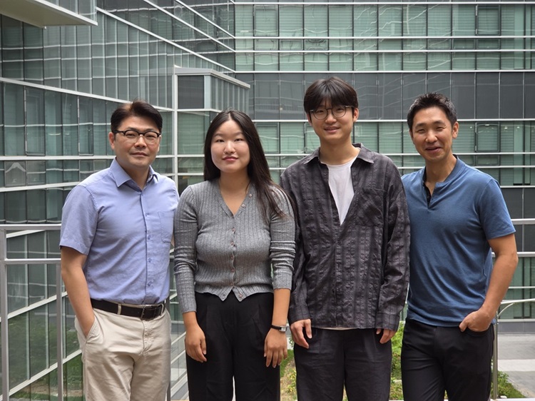
<(From left) Professor Dong-Wook Kim of Uijeongbu Eulji University Hospital Hematologic Malignancy Center, Professor Hongtae Kim of UNIST, Professor Chunghun Lim of KAIST, and Dr. Jumin Park of KAIST>
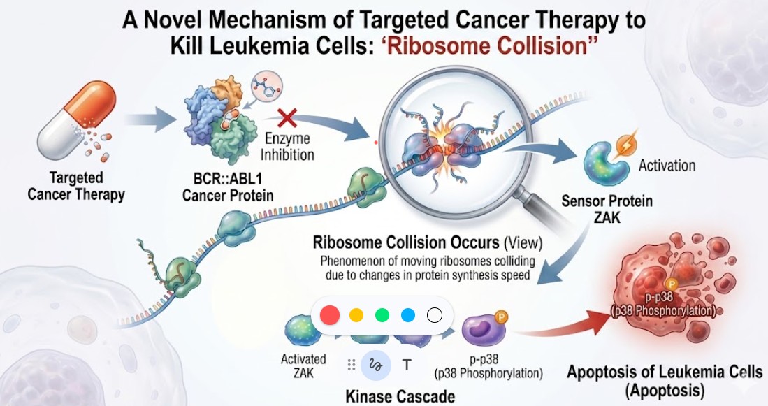
The real reason why anticancer drugs kill cancer cells has been revealed. KAIST research team has identified that targeted anticancer therapies do not simply block cancer proteins, but rather shut down the "protein factories" inside the cells, forcing them to undergo self-destruction. Consequently, the "two-faced protein" that plays a key role in this process is gaining attention as a breakthrough for treating patients with drug resistance.
KAIST announced on April 23rd that a joint research team—consisting of Professor Chunghun Lim from the Department of Biological Sciences at KAIST, Professor Dong-Wook Kim from the Hematologic Malignancy Center at Uijeongbu Eulji University Hospital, and Professor Hongtae Kim from UNIST —has identified a new molecular mechanism that regulates the response to anticancer drugs for Chronic Myeloid Leukemia (CML).
Chronic Myeloid Leukemia occurs when genetic abnormalities in hematopoietic stem cells produce an abnormal protein. This protein is known to be the primary cause of cancer cell proliferation by sending continuous growth signals to the cells. While targeted anticancer drugs that inhibit this protein are currently used as the standard treatment, there have been limitations, such as drug resistance or low treatment response in some patients.
The research team focused on the impact of anticancer drugs on the protein production process within the cell. As a result, they confirmed that when anticancer drugs are administered, the flow of ribosomes—the machines that create proteins—becomes tangled, leading to "ribosome collisions." This process induces intense stress inside the cell, ultimately leading the cancer cell to its death.
In particular, the research team identified the ZAK protein as the key sensor that detects these ribosome collisions and discovered that ZAK possesses "two faces" depending on the situation. Under normal conditions, it acts as an assistant, binding with AKT signals* to help cancer cells grow. However, once targeted anticancer treatment begins, it transforms into a sentinel that monitors ribosome collisions and triggers the death of the cancer cell. This marks the world's first proof that the same protein can perform diametrically opposite roles during cancer progression versus cancer treatment. *A key intracellular signaling pathway that regulates cell survival, growth, proliferation, metabolism, and migration.
<Clinical correlation between disease stage and ZAK expression in a Chronic Myeloid Leukemia patient cohort>
The research team verified this mechanism by analyzing cancer cells derived from actual leukemia patients. When drugs that increase ribosome collisions were used in combination, the anticancer effect improved significantly. Conversely, when ZAK function was impaired, the responsiveness to the anticancer drug decreased.
<Mechanism of ribosome collision and ZAK-dependent cancer cell death induced by Targeted Kinase Inhibitors (TKIs) in Chronic Myeloid Leukemia>
In other words, according to this study, drug-resistant patients are predicted to have decreased ZAK function or an insufficient ribosome stress response. This suggests that it is possible to predict treatment responses based on an individual patient's ZAK activation status and design customized combination therapy strategies.
This study is a significant achievement that presents the importance of the ribosome stress signaling pathway in the treatment of Chronic Myeloid Leukemia. It is expected to lead to the development of new combination therapies and enhance the effectiveness of targeted anticancer drugs. In particular, it offers new possibilities for patients struggling with drug resistance.
<Research Image (AI-generated)>
Professor Chunghun Lim stated, "This study shows how critical the process of the cell detecting abnormal protein synthesis and converting it into a death signal is for treatment." Dr. Jumin Park, the lead author, noted, "As we have confirmed that ribosome collision is a key switch determining cancer cell death, we plan to expand this research to various other types of cancer."
The results of this study, featuring Jumin Park of KAIST as the first author, were published online on March 30th in Leukemia, one of the most prestigious academic journals in the field of hematology.
Paper Title: BCR::ABL1 tyrosine kinase inhibitors induce ribosome collisions to activate ZAK-dependent ribotoxic stress and apoptosis in chronic myeloid leukemia
Authors: Jumin Park, Soo-Hyun Kim, Jongmin Park, Heeju Park, Hongtae Kim, Dong-Wook Kim & Chunghun Lim
DOI: https://doi.org/10.1038/s41375-026-02916-3
This research was conducted with support from the Suh Kyungbae Foundation, the Mid-career Researcher Support Program of the National Research Foundation of Korea, the Basic Research Lab Support Program, and the KAIST Settlement Project.
2026.04.23 View 431
Discovery of the Two-Faced Protein in Leukemia Treatment: A Clue to Overcoming Drug Resistance
<(From left) Professor Dong-Wook Kim of Uijeongbu Eulji University Hospital Hematologic Malignancy Center, Professor Hongtae Kim of UNIST, Professor Chunghun Lim of KAIST, and Dr. Jumin Park of KAIST>
The real reason why anticancer drugs kill cancer cells has been revealed. KAIST research team has identified that targeted anticancer therapies do not simply block cancer proteins, but rather shut down the "protein factories" inside the cells, forcing them to undergo self-destruction. Consequently, the "two-faced protein" that plays a key role in this process is gaining attention as a breakthrough for treating patients with drug resistance.
KAIST announced on April 23rd that a joint research team—consisting of Professor Chunghun Lim from the Department of Biological Sciences at KAIST, Professor Dong-Wook Kim from the Hematologic Malignancy Center at Uijeongbu Eulji University Hospital, and Professor Hongtae Kim from UNIST —has identified a new molecular mechanism that regulates the response to anticancer drugs for Chronic Myeloid Leukemia (CML).
Chronic Myeloid Leukemia occurs when genetic abnormalities in hematopoietic stem cells produce an abnormal protein. This protein is known to be the primary cause of cancer cell proliferation by sending continuous growth signals to the cells. While targeted anticancer drugs that inhibit this protein are currently used as the standard treatment, there have been limitations, such as drug resistance or low treatment response in some patients.
The research team focused on the impact of anticancer drugs on the protein production process within the cell. As a result, they confirmed that when anticancer drugs are administered, the flow of ribosomes—the machines that create proteins—becomes tangled, leading to "ribosome collisions." This process induces intense stress inside the cell, ultimately leading the cancer cell to its death.
In particular, the research team identified the ZAK protein as the key sensor that detects these ribosome collisions and discovered that ZAK possesses "two faces" depending on the situation. Under normal conditions, it acts as an assistant, binding with AKT signals* to help cancer cells grow. However, once targeted anticancer treatment begins, it transforms into a sentinel that monitors ribosome collisions and triggers the death of the cancer cell. This marks the world's first proof that the same protein can perform diametrically opposite roles during cancer progression versus cancer treatment. *A key intracellular signaling pathway that regulates cell survival, growth, proliferation, metabolism, and migration.
<Clinical correlation between disease stage and ZAK expression in a Chronic Myeloid Leukemia patient cohort>
The research team verified this mechanism by analyzing cancer cells derived from actual leukemia patients. When drugs that increase ribosome collisions were used in combination, the anticancer effect improved significantly. Conversely, when ZAK function was impaired, the responsiveness to the anticancer drug decreased.
<Mechanism of ribosome collision and ZAK-dependent cancer cell death induced by Targeted Kinase Inhibitors (TKIs) in Chronic Myeloid Leukemia>
In other words, according to this study, drug-resistant patients are predicted to have decreased ZAK function or an insufficient ribosome stress response. This suggests that it is possible to predict treatment responses based on an individual patient's ZAK activation status and design customized combination therapy strategies.
This study is a significant achievement that presents the importance of the ribosome stress signaling pathway in the treatment of Chronic Myeloid Leukemia. It is expected to lead to the development of new combination therapies and enhance the effectiveness of targeted anticancer drugs. In particular, it offers new possibilities for patients struggling with drug resistance.
<Research Image (AI-generated)>
Professor Chunghun Lim stated, "This study shows how critical the process of the cell detecting abnormal protein synthesis and converting it into a death signal is for treatment." Dr. Jumin Park, the lead author, noted, "As we have confirmed that ribosome collision is a key switch determining cancer cell death, we plan to expand this research to various other types of cancer."
The results of this study, featuring Jumin Park of KAIST as the first author, were published online on March 30th in Leukemia, one of the most prestigious academic journals in the field of hematology.
Paper Title: BCR::ABL1 tyrosine kinase inhibitors induce ribosome collisions to activate ZAK-dependent ribotoxic stress and apoptosis in chronic myeloid leukemia
Authors: Jumin Park, Soo-Hyun Kim, Jongmin Park, Heeju Park, Hongtae Kim, Dong-Wook Kim & Chunghun Lim
DOI: https://doi.org/10.1038/s41375-026-02916-3
This research was conducted with support from the Suh Kyungbae Foundation, the Mid-career Researcher Support Program of the National Research Foundation of Korea, the Basic Research Lab Support Program, and the KAIST Settlement Project.
2026.04.23 View 431 -
 KAIST, Flowers Have a “Biological Clock” That Times Blooming and Scent to Match Insects
Morning-blooming morning glories and flowers that release fragrance at night seem as if they know the time. A KAIST research team has uncovered, at the molecular level, the principle by which plants precisely control the timing of flower opening and scent emission through a “biological clock” aligned with insect behavior. This study suggests potential applications in technologies for controlling flowering time and fragrance.
KAIST (President Kwang Hyung Lee) announced on the 27th of March that a research team led by Professor Sang-Gyu Kim of the Department of Biological Sciences has discovered that genes regulated by the plant biological clock integratively control both the timing of flower opening and the circadian rhythm of scent emission.
Plants are known to have physiological processes regulated by a “biological clock” that allows them to perceive time according to a daily cycle. However, the exact process by which flowers open and how this process is connected to biological clock genes has not yet been fully elucidated.
The research team conducted their study using Nicotiana attenuata (coyote tobacco), a plant that opens its flowers widely at night and emits fragrance. Native to desert regions of Utah, USA, this plant is characterized by opening its flowers and releasing scent at night to attract nocturnal pollinators.
Inspired by such phenomena, the biologist Linnaeus proposed the idea of a “flower clock,” suggesting that if plants that bloom and close at different times were gathered together, one could tell the time based solely on their flowering states.
Previous studies mainly focused on analyzing changes in gene expression related to flower development, but research directly identifying the phenomenon of flower opening and the genes controlling it has been limited. To overcome these limitations, the research team analyzed mutants with altered biological clock genes and investigated, through molecular biological approaches, how flower opening and scent emission are regulated.
< A comparison of wild-type plants and COL5 (a specific circadian clock gene) mutant plants >
As a result, the team confirmed that specific biological clock genes play a key role in controlling both the timing of flower opening and the rhythm of scent emission. This demonstrates that plants make precise use of their biological clock to open flowers and attract pollinators at the most advantageous times for survival and reproduction.
< AI-generated image (the rhythmic behavior of coyote tobacco flowers regulated by the COL5 gene) >
This study is meaningful in that it presents a gene regulatory network controlling flower opening and scent emission from the perspective of the biological clock. It is also expected to provide important clues for understanding plants’ time-regulation strategies and their ecological interactions.
Professor Sang-Gyu Kim stated, “This study provides insight into how the plant biological clock links and regulates the timing of flower opening and scent emission,” adding, “It is expected to serve as an important foundation for understanding how plants optimize their reproductive strategies through interactions with the environment.”
This research, with Dr. Yuri Choi and Dr. Moonyoung Kang from the Department of Biological Sciences as co-first authors, was published on January 29 in the international journal The Plant Cell.
※ Paper title: ”CONSTANS-LIKE 5 facilitates flower opening and scent biosynthesis in Solanaceae” https://doi.org/10.1093/plcell/koag016
This research was supported by the National Research Foundation of Korea’s Synthetic Biology Core Technology Development Program, the Mid-career Researcher Program, and the Rural Development Administration’s Next-Generation Crop Breeding Technology Development Program.
2026.03.27 View 592
KAIST, Flowers Have a “Biological Clock” That Times Blooming and Scent to Match Insects
Morning-blooming morning glories and flowers that release fragrance at night seem as if they know the time. A KAIST research team has uncovered, at the molecular level, the principle by which plants precisely control the timing of flower opening and scent emission through a “biological clock” aligned with insect behavior. This study suggests potential applications in technologies for controlling flowering time and fragrance.
KAIST (President Kwang Hyung Lee) announced on the 27th of March that a research team led by Professor Sang-Gyu Kim of the Department of Biological Sciences has discovered that genes regulated by the plant biological clock integratively control both the timing of flower opening and the circadian rhythm of scent emission.
Plants are known to have physiological processes regulated by a “biological clock” that allows them to perceive time according to a daily cycle. However, the exact process by which flowers open and how this process is connected to biological clock genes has not yet been fully elucidated.
The research team conducted their study using Nicotiana attenuata (coyote tobacco), a plant that opens its flowers widely at night and emits fragrance. Native to desert regions of Utah, USA, this plant is characterized by opening its flowers and releasing scent at night to attract nocturnal pollinators.
Inspired by such phenomena, the biologist Linnaeus proposed the idea of a “flower clock,” suggesting that if plants that bloom and close at different times were gathered together, one could tell the time based solely on their flowering states.
Previous studies mainly focused on analyzing changes in gene expression related to flower development, but research directly identifying the phenomenon of flower opening and the genes controlling it has been limited. To overcome these limitations, the research team analyzed mutants with altered biological clock genes and investigated, through molecular biological approaches, how flower opening and scent emission are regulated.
< A comparison of wild-type plants and COL5 (a specific circadian clock gene) mutant plants >
As a result, the team confirmed that specific biological clock genes play a key role in controlling both the timing of flower opening and the rhythm of scent emission. This demonstrates that plants make precise use of their biological clock to open flowers and attract pollinators at the most advantageous times for survival and reproduction.
< AI-generated image (the rhythmic behavior of coyote tobacco flowers regulated by the COL5 gene) >
This study is meaningful in that it presents a gene regulatory network controlling flower opening and scent emission from the perspective of the biological clock. It is also expected to provide important clues for understanding plants’ time-regulation strategies and their ecological interactions.
Professor Sang-Gyu Kim stated, “This study provides insight into how the plant biological clock links and regulates the timing of flower opening and scent emission,” adding, “It is expected to serve as an important foundation for understanding how plants optimize their reproductive strategies through interactions with the environment.”
This research, with Dr. Yuri Choi and Dr. Moonyoung Kang from the Department of Biological Sciences as co-first authors, was published on January 29 in the international journal The Plant Cell.
※ Paper title: ”CONSTANS-LIKE 5 facilitates flower opening and scent biosynthesis in Solanaceae” https://doi.org/10.1093/plcell/koag016
This research was supported by the National Research Foundation of Korea’s Synthetic Biology Core Technology Development Program, the Mid-career Researcher Program, and the Rural Development Administration’s Next-Generation Crop Breeding Technology Development Program.
2026.03.27 View 592 -
 AI-Engineered "Nasal Spray Antiviral Platform" Developed to Block Flu and COVID-19
<(From Left) Professor Hyun Jung Chung, Professor Ho Min Kim, Professor Ji Eun Oh>
<(From Left) Dr. Seungju Yang, Dr. Jeongwon Yun, Ph.D candidate Jae Hyuk Kwon>
Respiratory viruses that have diverse strains and mutate rapidly, such as influenza and COVID-19, are difficult to block perfectly with vaccines alone. To solve this problem, KAIST's research team has successfully developed a nasal (intranasal) antiviral platform using AI technology to overcome the existing limitations of interferon-lambda treatments—namely, being "weak against heat and disappearing quickly from the nasal mucosa."
KAIST announced on December 15th that a joint research team—consisting of Professor Ho Min Ktim and Professor Hyun Jung Chung from the Department of Biological Sciences, and Professor Ji Eun Oh from the Graduate School of Medical Science and Engineering used AI to stably redesign the interferon-lambda protein and combined it with a delivery technology that ensures effective diffusion and long-term retention in the nasal mucosa, thereby implementing a universal prevention technology for various respiratory viruses.
Interferon-lambda is an innate immune protein produced by the body to block viral infections, playing a crucial role in stopping respiratory viruses like the common cold, flu, and COVID-19. However, when formulated as a treatment for nasal administration, its actual efficacy was limited by its vulnerability to heat, degrading enzymes, mucus, and ciliary motion.
The research team used AI protein design technology to precisely reinforce the structural weaknesses of interferon-lambda.
First, they significantly increased stability by changing the loose "loop" structures of the protein—which were prone to instability—into rigid "helix" structures that lock in place like a firm spring.
Additionally, to prevent "aggregation" (proteins sticking together to form lumps), they applied "surface engineering" to make the surface more water-compatible. They also introduced "glycoengineering," adding sugar chain (glycan) structures to the protein surface to make it even more robust and stable.
As a result, the newly produced interferon-lambda showed a massive improvement in stability, surviving for two weeks 50℃ and demonstrated the ability to diffuse rapidly even through thick nasal mucus.
The research team further protected the protein by encapsulating it in microscopic "nanoliposomes" and coated the surface with "low-molecular-weight chitosan." This significantly enhanced "mucoadhesion," allowing the treatment to stick to the nasal lining for an extended period.
When this delivery platform was applied to animal models infected with influenza, a powerful inhibitory effect was confirmed, with the virus level in the nasal cavity decreasing by more than 85%.
This technology is a mucosal immune platform that can block viral infections in their early stages simply by spraying it into the nose. It is expected to be a new therapeutic strategy that can respond quickly not only to seasonal flu but also to unexpected new or mutant viruses.
Professor Ho Min Kim stated, "Through AI-based protein design and mucosal delivery technology, we have simultaneously overcome the stability and retention time limitations of existing interferon-lambda treatments. This platform, which is stable at high temperatures and stays in the mucosa for a long time, is an innovative technology that can be used even in developing countries lacking strict cold-chain infrastructure. It also has great scalability for developing various treatments and vaccines." He added, "This is a meaningful achievement resulting from multidisciplinary convergence research, covering everything from AI protein design to drug delivery optimization and immune evaluation through infection models."
This research involved Dr. Jeongwon Yun from the KAIST InnoCORE (AI-Co-Research & Eudcation for innovative Drug Institute, AI-CRED Institute) Dr. Seungju Yang from the Department of Biological Sciences, and PhD student Jae Hyuk Kwon from the Graduate School of Medical Science and Engineering as co-first authors. The results were published consecutively in the renowned international journals Advanced Science (Nov 20) and Biomaterials Research (Nov 21).
Paper 1: Computational Design and Glycoengineering of Interferon-Lambda for Nasal Prophylaxis against Respiratory Viruses, Advanced Science, DOI: 10.1002/advs.202506764
Paper 2: Intranasal Nanoliposomes Delivering Interferon Lambda with Enhanced Mucosal Retention as an Antiviral, Biomaterials Research, DOI: 10.34133/bmr.0287
This research was conducted with support from the KAIST InnoCORE Program, Mid-Career Researcher Support Program and the Bio-Medical Technology Development Program through the National Research Foundation of Korea (NRF), Healthcare Technology R&D Project through the Korea Health Industry Development Institute (KHIDI), the KAIST Convergence Research Institute Operation Program, and the Institute for Basic Science (IBS).
2025.12.15 View 2414
AI-Engineered "Nasal Spray Antiviral Platform" Developed to Block Flu and COVID-19
<(From Left) Professor Hyun Jung Chung, Professor Ho Min Kim, Professor Ji Eun Oh>
<(From Left) Dr. Seungju Yang, Dr. Jeongwon Yun, Ph.D candidate Jae Hyuk Kwon>
Respiratory viruses that have diverse strains and mutate rapidly, such as influenza and COVID-19, are difficult to block perfectly with vaccines alone. To solve this problem, KAIST's research team has successfully developed a nasal (intranasal) antiviral platform using AI technology to overcome the existing limitations of interferon-lambda treatments—namely, being "weak against heat and disappearing quickly from the nasal mucosa."
KAIST announced on December 15th that a joint research team—consisting of Professor Ho Min Ktim and Professor Hyun Jung Chung from the Department of Biological Sciences, and Professor Ji Eun Oh from the Graduate School of Medical Science and Engineering used AI to stably redesign the interferon-lambda protein and combined it with a delivery technology that ensures effective diffusion and long-term retention in the nasal mucosa, thereby implementing a universal prevention technology for various respiratory viruses.
Interferon-lambda is an innate immune protein produced by the body to block viral infections, playing a crucial role in stopping respiratory viruses like the common cold, flu, and COVID-19. However, when formulated as a treatment for nasal administration, its actual efficacy was limited by its vulnerability to heat, degrading enzymes, mucus, and ciliary motion.
The research team used AI protein design technology to precisely reinforce the structural weaknesses of interferon-lambda.
First, they significantly increased stability by changing the loose "loop" structures of the protein—which were prone to instability—into rigid "helix" structures that lock in place like a firm spring.
Additionally, to prevent "aggregation" (proteins sticking together to form lumps), they applied "surface engineering" to make the surface more water-compatible. They also introduced "glycoengineering," adding sugar chain (glycan) structures to the protein surface to make it even more robust and stable.
As a result, the newly produced interferon-lambda showed a massive improvement in stability, surviving for two weeks 50℃ and demonstrated the ability to diffuse rapidly even through thick nasal mucus.
The research team further protected the protein by encapsulating it in microscopic "nanoliposomes" and coated the surface with "low-molecular-weight chitosan." This significantly enhanced "mucoadhesion," allowing the treatment to stick to the nasal lining for an extended period.
When this delivery platform was applied to animal models infected with influenza, a powerful inhibitory effect was confirmed, with the virus level in the nasal cavity decreasing by more than 85%.
This technology is a mucosal immune platform that can block viral infections in their early stages simply by spraying it into the nose. It is expected to be a new therapeutic strategy that can respond quickly not only to seasonal flu but also to unexpected new or mutant viruses.
Professor Ho Min Kim stated, "Through AI-based protein design and mucosal delivery technology, we have simultaneously overcome the stability and retention time limitations of existing interferon-lambda treatments. This platform, which is stable at high temperatures and stays in the mucosa for a long time, is an innovative technology that can be used even in developing countries lacking strict cold-chain infrastructure. It also has great scalability for developing various treatments and vaccines." He added, "This is a meaningful achievement resulting from multidisciplinary convergence research, covering everything from AI protein design to drug delivery optimization and immune evaluation through infection models."
This research involved Dr. Jeongwon Yun from the KAIST InnoCORE (AI-Co-Research & Eudcation for innovative Drug Institute, AI-CRED Institute) Dr. Seungju Yang from the Department of Biological Sciences, and PhD student Jae Hyuk Kwon from the Graduate School of Medical Science and Engineering as co-first authors. The results were published consecutively in the renowned international journals Advanced Science (Nov 20) and Biomaterials Research (Nov 21).
Paper 1: Computational Design and Glycoengineering of Interferon-Lambda for Nasal Prophylaxis against Respiratory Viruses, Advanced Science, DOI: 10.1002/advs.202506764
Paper 2: Intranasal Nanoliposomes Delivering Interferon Lambda with Enhanced Mucosal Retention as an Antiviral, Biomaterials Research, DOI: 10.34133/bmr.0287
This research was conducted with support from the KAIST InnoCORE Program, Mid-Career Researcher Support Program and the Bio-Medical Technology Development Program through the National Research Foundation of Korea (NRF), Healthcare Technology R&D Project through the Korea Health Industry Development Institute (KHIDI), the KAIST Convergence Research Institute Operation Program, and the Institute for Basic Science (IBS).
2025.12.15 View 2414 -
 KAIST Discovers Role of Huntingtin Protein in Building the Cell Skeleton
<(From Left) Professor Ji-Joon Song, Ph.D candidate Jaesung Kim, Dr. Hyeongju Kim of KAIST’s Department of Biological Sciences>
Huntington’s disease is a rare genetic disorder and a representative neurodegenerative disease, characterized by loss of motor control, cognitive decline, and psychiatric problems. An international research team has discovered that the “huntingtin protein,” the causal protein of Huntington’s disease (whose mutations are the direct cause of the disease), also performs a new function: directly organizing the cytoskeleton, the fine structural framework inside cells. This discovery is expected to contribute not only to understanding the pathogenic mechanism of Huntington’s disease, but also to research on neurodevelopmental disorders such as Alzheimer’s disease and Parkinson’s disease, as well as muscle- or mobility-related diseases such as muscular dystrophy.
KAIST (President Kwang Hyung Lee) announced on September 30 that a research team led by Professor Ji-Joon Song of the Department of Biological Sciences, in collaboration with the Institute of Science and Technology Austria (ISTA), Sorbonne University/Paris Brain Institute, and the Swiss Federal Institute of Technology Lausanne (EPFL), has uncovered—through cryo-electron microscopy (cryo-EM) and cell biology methods—the structural principle by which the huntingtin protein arranges cytoskeletal microfilaments (F-actin) into bundles.
Until now, the huntingtin protein was known only to “use” the cytoskeleton, being involved in vesicle transport or microtubule-based transport. The team, however, demonstrated that huntingtin physically organizes the cytoskeleton itself. This study is considered the first in the world to prove this new role of the huntingtin protein at the molecular level.
The researchers confirmed that huntingtin binds directly to cytoskeletal microfilaments (F-actin), and that pairs of huntingtin proteins bundle the cytoskeleton into arrays at intervals of about 20 nanometers.
Such cytoskeletal bundles play a crucial role in the development of neural connectivity. Indeed, structural development of neurons was found to be impaired in nerve cells deficient in the huntingtin protein.
<Elucidation of the Mechanism of Cytoskeletal Microfilament Bundle Formation by Huntingtin Protein and Its Impact on Neuronal Development>
First author Jaesung Kim, a PhD candidate at KAIST, stated, “This study provides a new perspective for understanding the molecular mechanism of the huntingtin protein, the cause of an incurable disease that has long remained a mystery.”
Professor Ji-Joon Song of KAIST’s Department of Biological Sciences explained, “This achievement not only provides an important clue to understanding the pathogenic mechanism of Huntington’s disease, but is also expected to have a far-reaching impact on research into cytoskeleton-related diseases,” and added that “it opens new avenues for exploring the role of the huntingtin protein in diverse biological phenomena such as cell division, migration, and mechanical signal transduction.”
This research was conducted with Jaesung Kim (PhD candidate, KAIST), Hyeongju Kim (now at Harvard University), Rémi Carpentier (Paris Brain Institute), Mariacristina Capizzi (Paris Brain Institute), and others as co-first authors, and was published on September 19 in Science Advances, a sister journal of Science.
※ Paper title: “Structure of the Huntingtin F-actin complex reveals its role in cytoskeleton organization,” DOI: https://doi.org/10.1126/sciadv.adw4124※ Co-corresponding authors: Ji-Joon Song (KAIST), Florian Schur (ISTA), and Sandrine Humbert (Sorbonne University/Paris Brain Institute).
This research was supported by the Ministry of Health and Welfare’s Global Research Collaboration Program (Korea–Switzerland Biohealth International Joint Research) and the Korea–Austria Cooperation Program.
2025.09.30 View 3108
KAIST Discovers Role of Huntingtin Protein in Building the Cell Skeleton
<(From Left) Professor Ji-Joon Song, Ph.D candidate Jaesung Kim, Dr. Hyeongju Kim of KAIST’s Department of Biological Sciences>
Huntington’s disease is a rare genetic disorder and a representative neurodegenerative disease, characterized by loss of motor control, cognitive decline, and psychiatric problems. An international research team has discovered that the “huntingtin protein,” the causal protein of Huntington’s disease (whose mutations are the direct cause of the disease), also performs a new function: directly organizing the cytoskeleton, the fine structural framework inside cells. This discovery is expected to contribute not only to understanding the pathogenic mechanism of Huntington’s disease, but also to research on neurodevelopmental disorders such as Alzheimer’s disease and Parkinson’s disease, as well as muscle- or mobility-related diseases such as muscular dystrophy.
KAIST (President Kwang Hyung Lee) announced on September 30 that a research team led by Professor Ji-Joon Song of the Department of Biological Sciences, in collaboration with the Institute of Science and Technology Austria (ISTA), Sorbonne University/Paris Brain Institute, and the Swiss Federal Institute of Technology Lausanne (EPFL), has uncovered—through cryo-electron microscopy (cryo-EM) and cell biology methods—the structural principle by which the huntingtin protein arranges cytoskeletal microfilaments (F-actin) into bundles.
Until now, the huntingtin protein was known only to “use” the cytoskeleton, being involved in vesicle transport or microtubule-based transport. The team, however, demonstrated that huntingtin physically organizes the cytoskeleton itself. This study is considered the first in the world to prove this new role of the huntingtin protein at the molecular level.
The researchers confirmed that huntingtin binds directly to cytoskeletal microfilaments (F-actin), and that pairs of huntingtin proteins bundle the cytoskeleton into arrays at intervals of about 20 nanometers.
Such cytoskeletal bundles play a crucial role in the development of neural connectivity. Indeed, structural development of neurons was found to be impaired in nerve cells deficient in the huntingtin protein.
<Elucidation of the Mechanism of Cytoskeletal Microfilament Bundle Formation by Huntingtin Protein and Its Impact on Neuronal Development>
First author Jaesung Kim, a PhD candidate at KAIST, stated, “This study provides a new perspective for understanding the molecular mechanism of the huntingtin protein, the cause of an incurable disease that has long remained a mystery.”
Professor Ji-Joon Song of KAIST’s Department of Biological Sciences explained, “This achievement not only provides an important clue to understanding the pathogenic mechanism of Huntington’s disease, but is also expected to have a far-reaching impact on research into cytoskeleton-related diseases,” and added that “it opens new avenues for exploring the role of the huntingtin protein in diverse biological phenomena such as cell division, migration, and mechanical signal transduction.”
This research was conducted with Jaesung Kim (PhD candidate, KAIST), Hyeongju Kim (now at Harvard University), Rémi Carpentier (Paris Brain Institute), Mariacristina Capizzi (Paris Brain Institute), and others as co-first authors, and was published on September 19 in Science Advances, a sister journal of Science.
※ Paper title: “Structure of the Huntingtin F-actin complex reveals its role in cytoskeleton organization,” DOI: https://doi.org/10.1126/sciadv.adw4124※ Co-corresponding authors: Ji-Joon Song (KAIST), Florian Schur (ISTA), and Sandrine Humbert (Sorbonne University/Paris Brain Institute).
This research was supported by the Ministry of Health and Welfare’s Global Research Collaboration Program (Korea–Switzerland Biohealth International Joint Research) and the Korea–Austria Cooperation Program.
2025.09.30 View 3108 -
 KAIST team links early life epigenetic memory to adult brain inflammation
<(From left) Professor Won-Suk Chung, Ph.D. Ph.D candidate Hyeonji Park Dr. Seongwan Park, Professor Inkyung Jung>
Why do some people remain healthy through childhood yet become more vulnerable to brain disorders such as dementia later in life? A KAIST (President Kwang Hyung Lee) -led team has uncovered a key part of the answer: a developmental ‘switch’ in astrocytes—the brain’s most abundant support cells that shapes how strongly the brain’s immune system reacts in adulthood. The study identifies a gene, NR3C1 (encoding the glucocorticoid receptor), as a master regulator of this switch and shows how early-life epigenetic ‘memory’ can predispose the adult brain to excessive inflammation.
The work was carried out by a joint team led by Professor Inkyung Jung (Department of Biological Sciences, KAIST) and Associate Director Won-Suk Chung (Center for Vascular Research, Institute for Basic Science; Professor, KAIST Biological Sciences). Using mouse models, the researchers mapped gene-regulatory programs across multiple stages of astrocyte development and found that NR3C1 acts during a brief early-postnatal window to enforce long-term immune restraint.
<The schematic illustrates how the NR3C1 gene (glucocorticoid receptor) suppresses the immune response of astrocytes. In normal (control) astrocytes, NR3C1 binds to specific regulatory regions of DNA (nGRE) to inhibit the expression of immune-related genes, thereby maintaining brain homeostasis even under immune stimulation. In contrast, in NR3C1-deficient astrocytes (KO), this suppression is lost, leading to excessive activation of inflammation-related genes such as Gfap, Il6st, Stat2, and Cxcl10. As a result, in an autoimmune encephalomyelitis (EAE) model, pronounced neuroinflammation and clinical symptoms (paralysis and severe debilitation) are observed>
To build this map, the team combined state-of-the-art 3D epigenome profiling with RNA sequencing and chromatin accessibility analyses, capturing how DNA folds and which regulatory elements contact target genes. They identified 55 stage-specific transcription factors that guide astrocyte maturation; among them, NR3C1 emerged as the critical ‘switch’ in early life. Notably, deleting NR3C1 in astrocytes did not disrupt normal development. However, when the adult mice were challenged with an autoimmune model of multiple sclerosis, animals lacking astrocytic NR3C1 mounted exaggerated inflammatory responses and developed more severe disease.
Mechanistically, the study shows that early loss of NR3C1 epigenetically primes immune genes - keeping their regulatory elements open and ready - so that later in life these genes respond too strongly to inflammatory cues. In effect, NR3C1 serves as an early ‘brake’ that prevents over-activation of astrocyte immune programs in adulthood.
“This is the first demonstration that astrocyte immune functions are governed by epigenetic memory,” said Professor Won-Suk Chung. “Our findings offer new clues to the origins of degenerative brain disorders, including Alzheimer’s disease.”
“We reveal a temporal regulatory window in astrocyte development that can set the stage for disease vulnerability in adulthood,” added Professor Inkyung Jung. “Understanding the 3D genome logic behind these programs could open paths to therapies for immune-related brain disorders such as multiple sclerosis.”
<The figure shows the three-dimensional genome structure of astrocytes at specific gene loci, illustrating how NR3C1 regulates their expression. In normal cells, NR3C1 binds to DNA and maintains the chromatin in a closed state, thereby preventing unnecessary activation between distal regulatory elements (enhancers) and gene promoters. In contrast, when NR3C1 is absent, the chromatin becomes open, creating a state in which enhancers and genes can be more easily activated. As a result, genes such as Mxi1 are overexpressed, triggering inflammatory responses. This clearly demonstrates that NR3C1 plays an essential role in maintaining immune homeostasis by stabilizing three-dimensional gene regulatory mechanisms.>
The results of this study were published online on September 22 in the international journal Nature Communications (IF 15.7), with Dr. Seongwan Park and PhD student Hyeonji Park of KAIST’s Department of Biological Sciences as co-first authors.
※ Paper title: “NR3C1-mediated epigenetic regulation suppresses astrocytic immune responses in mice,” DOI: https://www.nature.com/articles/s41467-025-64088-5
In addition, on September 17, the journal published a commentary article introducing this research: https://www.nature.com/articles/s41467-025-64102-w
This research was supported by the Suh Kyungbae Science Foundation, the Ministry of Health and Welfare, the Ministry of Science and ICT, and IBS.
Glossary - Epigenetic priming: preparing genes for rapid future activation by altering chromatin without changing DNA sequence
2025.09.25 View 3643
KAIST team links early life epigenetic memory to adult brain inflammation
<(From left) Professor Won-Suk Chung, Ph.D. Ph.D candidate Hyeonji Park Dr. Seongwan Park, Professor Inkyung Jung>
Why do some people remain healthy through childhood yet become more vulnerable to brain disorders such as dementia later in life? A KAIST (President Kwang Hyung Lee) -led team has uncovered a key part of the answer: a developmental ‘switch’ in astrocytes—the brain’s most abundant support cells that shapes how strongly the brain’s immune system reacts in adulthood. The study identifies a gene, NR3C1 (encoding the glucocorticoid receptor), as a master regulator of this switch and shows how early-life epigenetic ‘memory’ can predispose the adult brain to excessive inflammation.
The work was carried out by a joint team led by Professor Inkyung Jung (Department of Biological Sciences, KAIST) and Associate Director Won-Suk Chung (Center for Vascular Research, Institute for Basic Science; Professor, KAIST Biological Sciences). Using mouse models, the researchers mapped gene-regulatory programs across multiple stages of astrocyte development and found that NR3C1 acts during a brief early-postnatal window to enforce long-term immune restraint.
<The schematic illustrates how the NR3C1 gene (glucocorticoid receptor) suppresses the immune response of astrocytes. In normal (control) astrocytes, NR3C1 binds to specific regulatory regions of DNA (nGRE) to inhibit the expression of immune-related genes, thereby maintaining brain homeostasis even under immune stimulation. In contrast, in NR3C1-deficient astrocytes (KO), this suppression is lost, leading to excessive activation of inflammation-related genes such as Gfap, Il6st, Stat2, and Cxcl10. As a result, in an autoimmune encephalomyelitis (EAE) model, pronounced neuroinflammation and clinical symptoms (paralysis and severe debilitation) are observed>
To build this map, the team combined state-of-the-art 3D epigenome profiling with RNA sequencing and chromatin accessibility analyses, capturing how DNA folds and which regulatory elements contact target genes. They identified 55 stage-specific transcription factors that guide astrocyte maturation; among them, NR3C1 emerged as the critical ‘switch’ in early life. Notably, deleting NR3C1 in astrocytes did not disrupt normal development. However, when the adult mice were challenged with an autoimmune model of multiple sclerosis, animals lacking astrocytic NR3C1 mounted exaggerated inflammatory responses and developed more severe disease.
Mechanistically, the study shows that early loss of NR3C1 epigenetically primes immune genes - keeping their regulatory elements open and ready - so that later in life these genes respond too strongly to inflammatory cues. In effect, NR3C1 serves as an early ‘brake’ that prevents over-activation of astrocyte immune programs in adulthood.
“This is the first demonstration that astrocyte immune functions are governed by epigenetic memory,” said Professor Won-Suk Chung. “Our findings offer new clues to the origins of degenerative brain disorders, including Alzheimer’s disease.”
“We reveal a temporal regulatory window in astrocyte development that can set the stage for disease vulnerability in adulthood,” added Professor Inkyung Jung. “Understanding the 3D genome logic behind these programs could open paths to therapies for immune-related brain disorders such as multiple sclerosis.”
<The figure shows the three-dimensional genome structure of astrocytes at specific gene loci, illustrating how NR3C1 regulates their expression. In normal cells, NR3C1 binds to DNA and maintains the chromatin in a closed state, thereby preventing unnecessary activation between distal regulatory elements (enhancers) and gene promoters. In contrast, when NR3C1 is absent, the chromatin becomes open, creating a state in which enhancers and genes can be more easily activated. As a result, genes such as Mxi1 are overexpressed, triggering inflammatory responses. This clearly demonstrates that NR3C1 plays an essential role in maintaining immune homeostasis by stabilizing three-dimensional gene regulatory mechanisms.>
The results of this study were published online on September 22 in the international journal Nature Communications (IF 15.7), with Dr. Seongwan Park and PhD student Hyeonji Park of KAIST’s Department of Biological Sciences as co-first authors.
※ Paper title: “NR3C1-mediated epigenetic regulation suppresses astrocytic immune responses in mice,” DOI: https://www.nature.com/articles/s41467-025-64088-5
In addition, on September 17, the journal published a commentary article introducing this research: https://www.nature.com/articles/s41467-025-64102-w
This research was supported by the Suh Kyungbae Science Foundation, the Ministry of Health and Welfare, the Ministry of Science and ICT, and IBS.
Glossary - Epigenetic priming: preparing genes for rapid future activation by altering chromatin without changing DNA sequence
2025.09.25 View 3643 -
 KAIST Identifies Key to Slowing Aging via RNA Regulation... Unlocks Mechanism for Longevity
As aging progresses, the quality of DNA and proteins inside cells declines, known to be the cause of various degenerative diseases. However, the connection between aging and RNA has remained largely unexplored. Now, a Korean research team has discovered that a ribosome-associated quality control factor—PELOTA, a protein essential for eliminating abnormal mRNA—plays a central role in slowing aging and promoting longevity. This breakthrough is expected to provide a new direction for future therapeutic strategies targeting human aging and neurodegenerative diseases.
KAIST (President Kwang Hyung Lee) announced that a joint research team—led by Professor Seung-Jae V. Lee of the Department of Biological Sciences at KAIST and the Research Center for RNA-mediated Healthy Longevity, Professor Jinsoo Seo of Yonsei University (President Dong-Sup Yoon), and Professor Kwang-Pyo Lee of the Korea Research Institute of Bioscience and Biotechnology (KRIBB, President Suk Yoon Kwon) under the National Research Council of Science & Technology (NST, Chairman Yeung-Shik Kim—has discovered that the protein ‘PELOTA*’, which plays a key role in ribosome-associated quality control, regulates the pace of aging.
*PELOTA: A key protein in maintaining cellular translational homeostasis, responsible for detecting and resolving errors during mRNA translation by ribosomes.
Until now, RNA—particularly mRNA—has generally been regarded as a transient intermediary in protein synthesis. Its instability made it difficult to study quantitatively or track over time, leaving its physiological and functional roles relatively understudied compared to DNA.
Using C. elegans (a nematode widely used in aging research due to its short lifespan), the researchers first discovered that the ribosome-associated quality control factor PELOTA is essential for longevity. In particular, when PELOTA was overexpressed in normal nematodes, their lifespan was extended, suggesting that ribosome-associated quality control mechanisms involved in removing abnormal mRNA are necessary for promoting longevity.
The study also revealed that the ribosome-associated quality control system simultaneously regulates both the mTOR signaling pathway—which senses nutrient status or growth signals to control cell growth, protein synthesis, and autophagy, and plays a key role in aging and energy metabolism—and the autophagy pathway, the cellular cleanup and recycling system through which cells break down and reuse unnecessary or damaged components.
When PELOTA was deficient, the mTOR pathway became abnormally activated, and autophagy was suppressed—accelerating aging. Conversely, activation of PELOTA inhibited mTOR and induced autophagy, thereby maintaining cellular homeostasis and extending lifespan.
Notably, this mechanism was found to be conserved in both mice and humans. The study also showed that the loss of PELOTA could contribute to muscle aging and Alzheimer’s disease, suggesting its relevance to age-related disorders.
These findings indicate that the study of PELOTA and ribosome-associated quality control could play an important role in developing therapeutic strategies for human aging and neurodegenerative diseases.
Professor Seung-Jae V. Lee of KAIST, who led the research, stated, “While the connection between quality control and aging has been well established at the DNA and protein levels, molecular evidence showing that RNA quality control also functionally contributes to lifespan regulation has been very limited.” He emphasized that the “study provides strong evidence that the removal of abnormal RNA is a central axis in the aging regulatory network.”
The study was published on August 5th in the prestigious journal PNAS (Proceedings of the National Academy of Sciences), with Dr. Jongsun Lee and Dr. Eun Ji Kim of KAIST, Dr. Bora Lee of KRIBB, and Dr. Hyein Lee of Yonsei University as co-first authors.
※ Title: Pelota-mediated ribosome-associated quality control counteracts aging and age-associated pathologies across species ※ DOI: https://doi.org/10.1073/pnas.2505217122
This research was supported by the Global Leader Research Project of the National Research Foundation of Korea.
2025.08.18 View 4709
KAIST Identifies Key to Slowing Aging via RNA Regulation... Unlocks Mechanism for Longevity
As aging progresses, the quality of DNA and proteins inside cells declines, known to be the cause of various degenerative diseases. However, the connection between aging and RNA has remained largely unexplored. Now, a Korean research team has discovered that a ribosome-associated quality control factor—PELOTA, a protein essential for eliminating abnormal mRNA—plays a central role in slowing aging and promoting longevity. This breakthrough is expected to provide a new direction for future therapeutic strategies targeting human aging and neurodegenerative diseases.
KAIST (President Kwang Hyung Lee) announced that a joint research team—led by Professor Seung-Jae V. Lee of the Department of Biological Sciences at KAIST and the Research Center for RNA-mediated Healthy Longevity, Professor Jinsoo Seo of Yonsei University (President Dong-Sup Yoon), and Professor Kwang-Pyo Lee of the Korea Research Institute of Bioscience and Biotechnology (KRIBB, President Suk Yoon Kwon) under the National Research Council of Science & Technology (NST, Chairman Yeung-Shik Kim—has discovered that the protein ‘PELOTA*’, which plays a key role in ribosome-associated quality control, regulates the pace of aging.
*PELOTA: A key protein in maintaining cellular translational homeostasis, responsible for detecting and resolving errors during mRNA translation by ribosomes.
Until now, RNA—particularly mRNA—has generally been regarded as a transient intermediary in protein synthesis. Its instability made it difficult to study quantitatively or track over time, leaving its physiological and functional roles relatively understudied compared to DNA.
Using C. elegans (a nematode widely used in aging research due to its short lifespan), the researchers first discovered that the ribosome-associated quality control factor PELOTA is essential for longevity. In particular, when PELOTA was overexpressed in normal nematodes, their lifespan was extended, suggesting that ribosome-associated quality control mechanisms involved in removing abnormal mRNA are necessary for promoting longevity.
The study also revealed that the ribosome-associated quality control system simultaneously regulates both the mTOR signaling pathway—which senses nutrient status or growth signals to control cell growth, protein synthesis, and autophagy, and plays a key role in aging and energy metabolism—and the autophagy pathway, the cellular cleanup and recycling system through which cells break down and reuse unnecessary or damaged components.
When PELOTA was deficient, the mTOR pathway became abnormally activated, and autophagy was suppressed—accelerating aging. Conversely, activation of PELOTA inhibited mTOR and induced autophagy, thereby maintaining cellular homeostasis and extending lifespan.
Notably, this mechanism was found to be conserved in both mice and humans. The study also showed that the loss of PELOTA could contribute to muscle aging and Alzheimer’s disease, suggesting its relevance to age-related disorders.
These findings indicate that the study of PELOTA and ribosome-associated quality control could play an important role in developing therapeutic strategies for human aging and neurodegenerative diseases.
Professor Seung-Jae V. Lee of KAIST, who led the research, stated, “While the connection between quality control and aging has been well established at the DNA and protein levels, molecular evidence showing that RNA quality control also functionally contributes to lifespan regulation has been very limited.” He emphasized that the “study provides strong evidence that the removal of abnormal RNA is a central axis in the aging regulatory network.”
The study was published on August 5th in the prestigious journal PNAS (Proceedings of the National Academy of Sciences), with Dr. Jongsun Lee and Dr. Eun Ji Kim of KAIST, Dr. Bora Lee of KRIBB, and Dr. Hyein Lee of Yonsei University as co-first authors.
※ Title: Pelota-mediated ribosome-associated quality control counteracts aging and age-associated pathologies across species ※ DOI: https://doi.org/10.1073/pnas.2505217122
This research was supported by the Global Leader Research Project of the National Research Foundation of Korea.
2025.08.18 View 4709 -
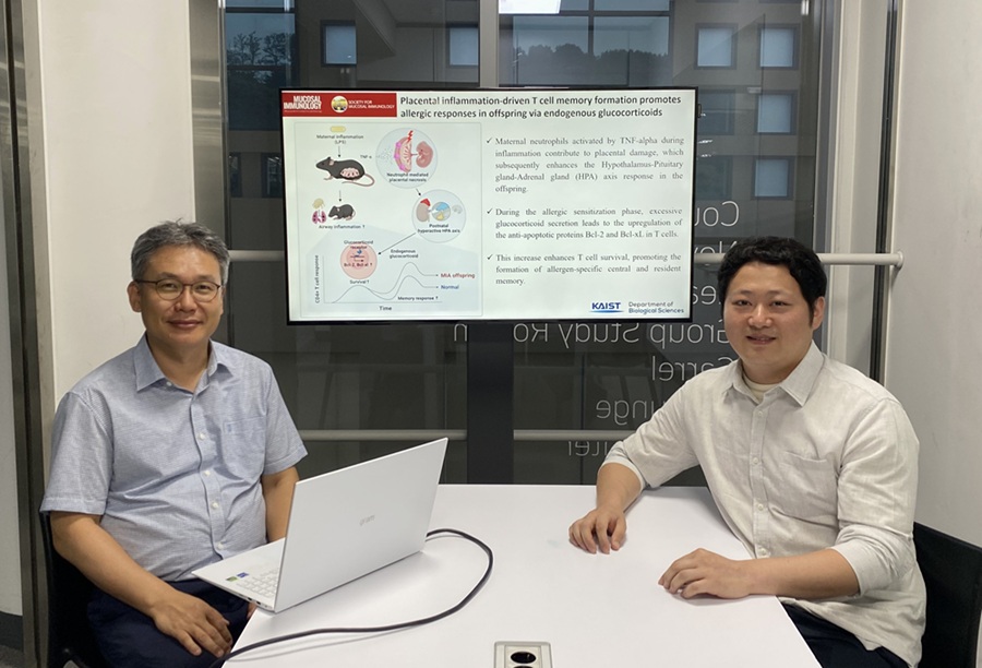 KAIST Reveals Placental Inflammation as the Cause of Allergies such as Pediatric Asthma
<(From left)Professor Heung-kyu Lee from the Department of Biological Sciences, Dr.Myeong Seung Kwon from the Graduate School of Medical Science>
It is already well-known that when a mother experiences inflammation during pregnancy, her child is more likely to develop allergic diseases. Recently, a KAIST research team became the first in the world to discover that inflammation within the placenta affects the fetus's immune system, leading to the child exhibiting excessive allergic reactions after birth. This study presents a new possibility for the early prediction and prevention of allergic diseases such as pediatric asthma.
KAIST (President Kwang Hyung Lee) announced on the 4th of August that a research team led by Professor Heung-kyu Lee from the Department of Biological Sciences found that inflammation occurring during pregnancy affects the fetus's stress response regulation system through the placenta. As a result, the survival and memory differentiation of T cells (key cells in the adaptive immune system) increase, which can lead to stronger allergic reactions in the child after birth.
The research team proved this through experiments on mice that had excessive inflammation induced during pregnancy. First, they injected the toxin component 'LPS (lipopolysaccharide),' a substance known to be a representative material that induces an inflammatory response in the immune system, into the mice to cause an inflammatory response in their bodies, which also caused inflammation in the placenta.
It was confirmed that the placental tissue, due to the inflammatory response, increased a signaling substance called 'Tumor Necrosis Factor-alpha (TNF-α),' and this substance activated immune cells called 'neutrophils*', causing inflammatory damage to the placenta. *Neutrophils: The most abundant type of white blood cells in our bodies (40-75%), playing an important role in innate immunity and killing invading bacteria and fungi.
This damage modulated postnatal offspring stress response, leading to a large secretion of stress hormone (glucocorticoid). As a result, the offspring's T cells, which are responsible for immune memory, survived longer and had stronger memory functions.
In particular, the memory T cells created through this process caused excessive allergic reactions when repeatedly exposed to antigens after birth. Specifically, when house dust mite 'allergens' were exposed to the airways of mice, a strong eosinophilic inflammatory response and excessive immune activation were observed, with an increase in immune cells important for allergy and asthma reactions.
Professor Heung Kyu Lee stated, "This study is the first in the world to identify how a mother's inflammatory response during pregnancy affects the fetus's allergic immune system through the placenta." He added, "This will be an important scientific basis for developing biomarkers for early prediction and establishing prevention strategies for pediatric allergic diseases."
The first author of this study is Dr. Myeong Seung Kwon from the KAIST Graduate School of Medical Science (currently a clinical fellow of gynecological oncology at Konyang University Hospital's Department of Obstetrics and Gynecology), and the research results were published in the authoritative journal in the field of mucosal immunology, 'Mucosal Immunology,' on July 1st. ※ Paper Title: Placental inflammation-driven T cell memory formation promotes allergic responses in offspring via endogenous glucocorticoids ※ DOI: https://doi.org/10.1016/j.mucimm.2025.06.006
This research was conducted as part of the Basic Science Research Program and the Bio-Medical Technology Development Program supported by the Ministry of Science and ICT and the National Research Foundation of Korea.
2025.08.03 View 2567
KAIST Reveals Placental Inflammation as the Cause of Allergies such as Pediatric Asthma
<(From left)Professor Heung-kyu Lee from the Department of Biological Sciences, Dr.Myeong Seung Kwon from the Graduate School of Medical Science>
It is already well-known that when a mother experiences inflammation during pregnancy, her child is more likely to develop allergic diseases. Recently, a KAIST research team became the first in the world to discover that inflammation within the placenta affects the fetus's immune system, leading to the child exhibiting excessive allergic reactions after birth. This study presents a new possibility for the early prediction and prevention of allergic diseases such as pediatric asthma.
KAIST (President Kwang Hyung Lee) announced on the 4th of August that a research team led by Professor Heung-kyu Lee from the Department of Biological Sciences found that inflammation occurring during pregnancy affects the fetus's stress response regulation system through the placenta. As a result, the survival and memory differentiation of T cells (key cells in the adaptive immune system) increase, which can lead to stronger allergic reactions in the child after birth.
The research team proved this through experiments on mice that had excessive inflammation induced during pregnancy. First, they injected the toxin component 'LPS (lipopolysaccharide),' a substance known to be a representative material that induces an inflammatory response in the immune system, into the mice to cause an inflammatory response in their bodies, which also caused inflammation in the placenta.
It was confirmed that the placental tissue, due to the inflammatory response, increased a signaling substance called 'Tumor Necrosis Factor-alpha (TNF-α),' and this substance activated immune cells called 'neutrophils*', causing inflammatory damage to the placenta. *Neutrophils: The most abundant type of white blood cells in our bodies (40-75%), playing an important role in innate immunity and killing invading bacteria and fungi.
This damage modulated postnatal offspring stress response, leading to a large secretion of stress hormone (glucocorticoid). As a result, the offspring's T cells, which are responsible for immune memory, survived longer and had stronger memory functions.
In particular, the memory T cells created through this process caused excessive allergic reactions when repeatedly exposed to antigens after birth. Specifically, when house dust mite 'allergens' were exposed to the airways of mice, a strong eosinophilic inflammatory response and excessive immune activation were observed, with an increase in immune cells important for allergy and asthma reactions.
Professor Heung Kyu Lee stated, "This study is the first in the world to identify how a mother's inflammatory response during pregnancy affects the fetus's allergic immune system through the placenta." He added, "This will be an important scientific basis for developing biomarkers for early prediction and establishing prevention strategies for pediatric allergic diseases."
The first author of this study is Dr. Myeong Seung Kwon from the KAIST Graduate School of Medical Science (currently a clinical fellow of gynecological oncology at Konyang University Hospital's Department of Obstetrics and Gynecology), and the research results were published in the authoritative journal in the field of mucosal immunology, 'Mucosal Immunology,' on July 1st. ※ Paper Title: Placental inflammation-driven T cell memory formation promotes allergic responses in offspring via endogenous glucocorticoids ※ DOI: https://doi.org/10.1016/j.mucimm.2025.06.006
This research was conducted as part of the Basic Science Research Program and the Bio-Medical Technology Development Program supported by the Ministry of Science and ICT and the National Research Foundation of Korea.
2025.08.03 View 2567 -
 Why Do Plants Attack Themselves? The Secret of Genetic Conflict Revealed
<Professor Ji-Joon Song of the KAIST Department of Biological Sciences>
Plants, with their unique immune systems, sometimes launch 'autoimmune responses' by mistakenly identifying their own protein structures as pathogens. In particular, 'hybrid necrosis,' a phenomenon where descendant plants fail to grow healthily and perish after cross-breeding different varieties, has long been a difficult challenge for botanists and agricultural researchers. In response, an international research team has successfully elucidated the mechanism inducing plant autoimmune responses and proposed a novel strategy for cultivar improvement that can predict and avoid these reactions.
Professor Ji-Joon Song's research team at KAIST, in collaboration with teams from the National University of Singapore (NUS) and the University of Oxford, announced on the 21st of July that they have elucidated the structure and function of the 'DM3' protein complex, which triggers plant autoimmune responses, using cryo-electron microscopy (Cryo-EM) technology.
This research is drawing attention because it identifies defects in protein structure as the cause of hybrid necrosis, which occurs due to an abnormal reaction of immune receptors during cross-breeding between plant hybrids.
This protein (DM3) is originally an enzyme involved in the plant's immune response, but problems arise when the structure of the DM3 protein is damaged in a specific protein combination called 'DANGEROUS MIX (DM)'.
Notably, one variant of DM3, the 'DM3Col-0' variant, forms a stable complex with six proteins and is recognized as normal, thus not triggering an immune response. In contrast, another 'DM3Hh-0' variant has improper binding between its six proteins, causing the plant to recognize it as an 'abnormal state' and trigger an immune alarm, leading to autoimmunity.
The research team visualized this structure using atomic-resolution cryo-electron microscopy (Cryo-EM) and revealed that the immune-inducing ability is not due to the enzymatic function of the DM3 protein, but rather to 'differences in protein binding affinity.'
<Figure 1. Mechanism of Plant Autoimmunity Triggered by the Collapse of the DM3 Protein Complex>
This demonstrates that plants can initiate an immune response by recognizing not only 'external pathogens' but also 'internal protein structures' when they undergo abnormal changes, treating them as if they were pathogens.
The study shows how sensitively the plant immune system changes and triggers autoimmune responses when genes are mixed and protein structures change during the cross-breeding of different plant varieties. It significantly advanced the understanding of genetic incompatibility that can occur during natural cross-breeding and cultivar improvement processes.
Dr. Gijeong Kim, the co-first author, stated, "Through international research collaboration, we presented a new perspective on understanding the plant immune system by leveraging the autoimmune phenomenon, completing a high-quality study that encompasses structural biochemistry, genetics, and cell biological experiments."
Professor Ji-Joon Song of the KAIST Department of Biological Sciences, who led the research, said, "The fact that the immune system can detect not only external pathogens but also structural abnormalities in its own proteins will set a new standard for plant biotechnology and crop breeding strategies. Cryo-electron microscopy-based structural analysis will be an important tool for understanding the essence of gene interactions."
This research, with Professor Ji-Joon Song and Professor Eunyoung Chae of the University of Oxford as co-corresponding authors, Dr. Gijeong Kim (currently a postdoctoral researcher at the University of Zurich) and Dr. Wei-Lin Wan of the National University of Singapore as co-first authors, and Ph.D candidate Nayun Kim, as the second author, was published on July 17th in Molecular Cell, a sister journal of the international academic journal Cell.
This research was supported by the KAIST Grand Challenge 30 project.
Article Title: Structural determinants of DANGEROUS MIX 3, an alpha/beta hydrolase that triggers NLR-mediated genetic incompatibility in plants DOI: https://doi.org/10.1016/j.molcel.2025.06.021
2025.07.21 View 4220
Why Do Plants Attack Themselves? The Secret of Genetic Conflict Revealed
<Professor Ji-Joon Song of the KAIST Department of Biological Sciences>
Plants, with their unique immune systems, sometimes launch 'autoimmune responses' by mistakenly identifying their own protein structures as pathogens. In particular, 'hybrid necrosis,' a phenomenon where descendant plants fail to grow healthily and perish after cross-breeding different varieties, has long been a difficult challenge for botanists and agricultural researchers. In response, an international research team has successfully elucidated the mechanism inducing plant autoimmune responses and proposed a novel strategy for cultivar improvement that can predict and avoid these reactions.
Professor Ji-Joon Song's research team at KAIST, in collaboration with teams from the National University of Singapore (NUS) and the University of Oxford, announced on the 21st of July that they have elucidated the structure and function of the 'DM3' protein complex, which triggers plant autoimmune responses, using cryo-electron microscopy (Cryo-EM) technology.
This research is drawing attention because it identifies defects in protein structure as the cause of hybrid necrosis, which occurs due to an abnormal reaction of immune receptors during cross-breeding between plant hybrids.
This protein (DM3) is originally an enzyme involved in the plant's immune response, but problems arise when the structure of the DM3 protein is damaged in a specific protein combination called 'DANGEROUS MIX (DM)'.
Notably, one variant of DM3, the 'DM3Col-0' variant, forms a stable complex with six proteins and is recognized as normal, thus not triggering an immune response. In contrast, another 'DM3Hh-0' variant has improper binding between its six proteins, causing the plant to recognize it as an 'abnormal state' and trigger an immune alarm, leading to autoimmunity.
The research team visualized this structure using atomic-resolution cryo-electron microscopy (Cryo-EM) and revealed that the immune-inducing ability is not due to the enzymatic function of the DM3 protein, but rather to 'differences in protein binding affinity.'
<Figure 1. Mechanism of Plant Autoimmunity Triggered by the Collapse of the DM3 Protein Complex>
This demonstrates that plants can initiate an immune response by recognizing not only 'external pathogens' but also 'internal protein structures' when they undergo abnormal changes, treating them as if they were pathogens.
The study shows how sensitively the plant immune system changes and triggers autoimmune responses when genes are mixed and protein structures change during the cross-breeding of different plant varieties. It significantly advanced the understanding of genetic incompatibility that can occur during natural cross-breeding and cultivar improvement processes.
Dr. Gijeong Kim, the co-first author, stated, "Through international research collaboration, we presented a new perspective on understanding the plant immune system by leveraging the autoimmune phenomenon, completing a high-quality study that encompasses structural biochemistry, genetics, and cell biological experiments."
Professor Ji-Joon Song of the KAIST Department of Biological Sciences, who led the research, said, "The fact that the immune system can detect not only external pathogens but also structural abnormalities in its own proteins will set a new standard for plant biotechnology and crop breeding strategies. Cryo-electron microscopy-based structural analysis will be an important tool for understanding the essence of gene interactions."
This research, with Professor Ji-Joon Song and Professor Eunyoung Chae of the University of Oxford as co-corresponding authors, Dr. Gijeong Kim (currently a postdoctoral researcher at the University of Zurich) and Dr. Wei-Lin Wan of the National University of Singapore as co-first authors, and Ph.D candidate Nayun Kim, as the second author, was published on July 17th in Molecular Cell, a sister journal of the international academic journal Cell.
This research was supported by the KAIST Grand Challenge 30 project.
Article Title: Structural determinants of DANGEROUS MIX 3, an alpha/beta hydrolase that triggers NLR-mediated genetic incompatibility in plants DOI: https://doi.org/10.1016/j.molcel.2025.06.021
2025.07.21 View 4220 -
 KAIST Shows That the Brain Can Distinguish Glucose: Clues to Treat Obesity and Diabetes
<(From left)Prof. Greg S.B Suh, Dr. Jieun Kim, Dr. Shinhye Kim, Researcher Wongyo Jeong)
“How does our brain distinguish glucose from the many nutrients absorbed in the gut?” Starting with this question, a KAIST research team has demonstrated that the brain can selectively recognize specific nutrients—particularly glucose—beyond simply detecting total calorie content. This study is expected to offer a new paradigm for appetite control and the treatment of metabolic diseases.
On the 9th, KAIST (President Kwang Hyung Lee) announced that Professor Greg S.B. Suh’s team in the Department of Biological Sciences, in collaboration with Professor Young-Gyun Park’s team (BarNeuro), Professor Seung-Hee Lee’s team (Department of Biological Sciences), and the Albert Einstein College of Medicine in New York, had identified the existence of a gut-brain circuit that allows animals in a hungry state to selectively detect and prefer glucose in the gut.
Organisms derive energy from various nutrients including sugars, proteins, and fats. Previous studies have shown that total caloric information in the gut suppresses hunger neurons in the hypothalamus to regulate appetite. However, the existence of a gut-brain circuit that specifically responds to glucose and corresponding brain cells had not been demonstrated until now.
In this study, the team successfully identified a “gut-brain circuit” that senses glucose—essential for brain function—and regulates food intake behavior for required nutrients.
They further proved, for the first time, that this circuit responds within seconds to not only hunger or external stimuli but also to specific caloric nutrients directly introduced into the small intestine, particularly D-glucose, through the activity of “CRF neurons*” in the brain’s hypothalamus.
*CRF neurons: These neurons secrete corticotropin-releasing factor (CRF) in the hypothalamus and are central to the hypothalamic-pituitary-adrenal (HPA) axis, the body’s core physiological system for responding to stress. CRF neurons are known to regulate neuroendocrine balance in response to stress stimuli.
Using optogenetics to precisely track neural activity in real time, the researchers injected various nutrients—D-glucose, L-glucose, amino acids, and fats—directly into the small intestines of mice and observed the results.
They discovered that among the CRF neurons located in the paraventricular nucleus (PVN)* of the hypothalamus, only those specific to D-glucose showed selective responses. These neurons did not respond—or showed inverse reactions—to other sugars or to proteins and fats. This is the first demonstration that single neurons in the brain can guide nutrient-specific responses depending on gut nutrient influx.
*PVN (Paraventricular Nucleus): A key nucleus within the hypothalamus responsible for maintaining bodily homeostasis.
The team also revealed that glucose-sensing signals in the small intestine are transmitted via the spinal cord to the dorsolateral parabrachial nucleus (PBNdl) of the brain, and from there to CRF neurons in the PVN. In contrast, signals for amino acids and fats are transmitted to the brain through the vagus nerve, a different pathway.
In optogenetic inhibition experiments, suppressing CRF neurons in fasting mice eliminated their preference for glucose, proving that this circuit is essential for glucose-specific nutrient preference.
This study was inspired by Professor Suh’s earlier research at NYU using fruit flies, where he identified “DH44 neurons” that selectively detect glucose and sugar in the gut. Based on the hypothesis that hypothalamic neurons in mammals would show similar functional responses to glucose, the current study was launched.
To test this hypothesis, Dr. Jineun Kim (KAIST Ph.D. graduate, now at Caltech) demonstrated during her doctoral research that hungry mice preferred glucose among various intragastrically infused nutrients and that CRF neurons exhibited rapid and specific responses.
Along with Wongyo Jung (KAIST B.S. graduate, now Ph.D. student at Caltech), they modeled and experimentally confirmed the critical role of CRF neurons. Dr. Shinhye Kim, through collaboration, revealed that specific spinal neurons play a key role in conveying intestinal nutrient information to the brain.
Dr. Jineun Kim and Dr. Shinhye Kim said, “This study started from a simple but fundamental question—‘How does the brain distinguish glucose from various nutrients absorbed in the gut?’ We have shown that spinal-based gut-brain circuits play a central role in energy metabolism and homeostasis by transmitting specific gut nutrient signals to the brain.”
Professor Suh added, “By identifying a gut-brain pathway specialized for glucose, this research offers a new therapeutic target for metabolic diseases such as obesity and diabetes. Our future research will explore similar circuits for sensing other essential nutrients like amino acids and fats and their interaction mechanisms.”
Ph.D. student Jineun Kim, Dr. Shinhye Kim, and student Wongyo Jung (co-first authors) contributed to this study, which was published online in the international journal Neuron on June 20, 2025.
※ Paper Title: Encoding the glucose identity by discrete hypothalamic neurons via the gut-brain axis ※ DOI: https://doi.org/10.1016/j.neuron.2025.05.024
This study was supported by the Samsung Science & Technology Foundation, the National Research Foundation of Korea (NRF) Leader Research Program, the POSCO Cheongam Science Fellowship, the Asan Foundation Biomedical Science Scholarship, the Institute for Basic Science (IBS), and the KAIST KAIX program.
2025.07.09 View 3743
KAIST Shows That the Brain Can Distinguish Glucose: Clues to Treat Obesity and Diabetes
<(From left)Prof. Greg S.B Suh, Dr. Jieun Kim, Dr. Shinhye Kim, Researcher Wongyo Jeong)
“How does our brain distinguish glucose from the many nutrients absorbed in the gut?” Starting with this question, a KAIST research team has demonstrated that the brain can selectively recognize specific nutrients—particularly glucose—beyond simply detecting total calorie content. This study is expected to offer a new paradigm for appetite control and the treatment of metabolic diseases.
On the 9th, KAIST (President Kwang Hyung Lee) announced that Professor Greg S.B. Suh’s team in the Department of Biological Sciences, in collaboration with Professor Young-Gyun Park’s team (BarNeuro), Professor Seung-Hee Lee’s team (Department of Biological Sciences), and the Albert Einstein College of Medicine in New York, had identified the existence of a gut-brain circuit that allows animals in a hungry state to selectively detect and prefer glucose in the gut.
Organisms derive energy from various nutrients including sugars, proteins, and fats. Previous studies have shown that total caloric information in the gut suppresses hunger neurons in the hypothalamus to regulate appetite. However, the existence of a gut-brain circuit that specifically responds to glucose and corresponding brain cells had not been demonstrated until now.
In this study, the team successfully identified a “gut-brain circuit” that senses glucose—essential for brain function—and regulates food intake behavior for required nutrients.
They further proved, for the first time, that this circuit responds within seconds to not only hunger or external stimuli but also to specific caloric nutrients directly introduced into the small intestine, particularly D-glucose, through the activity of “CRF neurons*” in the brain’s hypothalamus.
*CRF neurons: These neurons secrete corticotropin-releasing factor (CRF) in the hypothalamus and are central to the hypothalamic-pituitary-adrenal (HPA) axis, the body’s core physiological system for responding to stress. CRF neurons are known to regulate neuroendocrine balance in response to stress stimuli.
Using optogenetics to precisely track neural activity in real time, the researchers injected various nutrients—D-glucose, L-glucose, amino acids, and fats—directly into the small intestines of mice and observed the results.
They discovered that among the CRF neurons located in the paraventricular nucleus (PVN)* of the hypothalamus, only those specific to D-glucose showed selective responses. These neurons did not respond—or showed inverse reactions—to other sugars or to proteins and fats. This is the first demonstration that single neurons in the brain can guide nutrient-specific responses depending on gut nutrient influx.
*PVN (Paraventricular Nucleus): A key nucleus within the hypothalamus responsible for maintaining bodily homeostasis.
The team also revealed that glucose-sensing signals in the small intestine are transmitted via the spinal cord to the dorsolateral parabrachial nucleus (PBNdl) of the brain, and from there to CRF neurons in the PVN. In contrast, signals for amino acids and fats are transmitted to the brain through the vagus nerve, a different pathway.
In optogenetic inhibition experiments, suppressing CRF neurons in fasting mice eliminated their preference for glucose, proving that this circuit is essential for glucose-specific nutrient preference.
This study was inspired by Professor Suh’s earlier research at NYU using fruit flies, where he identified “DH44 neurons” that selectively detect glucose and sugar in the gut. Based on the hypothesis that hypothalamic neurons in mammals would show similar functional responses to glucose, the current study was launched.
To test this hypothesis, Dr. Jineun Kim (KAIST Ph.D. graduate, now at Caltech) demonstrated during her doctoral research that hungry mice preferred glucose among various intragastrically infused nutrients and that CRF neurons exhibited rapid and specific responses.
Along with Wongyo Jung (KAIST B.S. graduate, now Ph.D. student at Caltech), they modeled and experimentally confirmed the critical role of CRF neurons. Dr. Shinhye Kim, through collaboration, revealed that specific spinal neurons play a key role in conveying intestinal nutrient information to the brain.
Dr. Jineun Kim and Dr. Shinhye Kim said, “This study started from a simple but fundamental question—‘How does the brain distinguish glucose from various nutrients absorbed in the gut?’ We have shown that spinal-based gut-brain circuits play a central role in energy metabolism and homeostasis by transmitting specific gut nutrient signals to the brain.”
Professor Suh added, “By identifying a gut-brain pathway specialized for glucose, this research offers a new therapeutic target for metabolic diseases such as obesity and diabetes. Our future research will explore similar circuits for sensing other essential nutrients like amino acids and fats and their interaction mechanisms.”
Ph.D. student Jineun Kim, Dr. Shinhye Kim, and student Wongyo Jung (co-first authors) contributed to this study, which was published online in the international journal Neuron on June 20, 2025.
※ Paper Title: Encoding the glucose identity by discrete hypothalamic neurons via the gut-brain axis ※ DOI: https://doi.org/10.1016/j.neuron.2025.05.024
This study was supported by the Samsung Science & Technology Foundation, the National Research Foundation of Korea (NRF) Leader Research Program, the POSCO Cheongam Science Fellowship, the Asan Foundation Biomedical Science Scholarship, the Institute for Basic Science (IBS), and the KAIST KAIX program.
2025.07.09 View 3743 -
 KAIST Enhances Immunotherapy for Difficult-to-Treat Brain Tumors with Gut Microbiota
< Photo 1.(From left) Prof. Heung Kyu Lee, Department of Biological Sciences,
and Dr. Hyeon Cheol Kim>
Advanced treatments, known as immunotherapies that activate T cells—our body's immune cells—to eliminate cancer cells, have shown limited efficacy as standalone therapies for glioblastoma, the most lethal form of brain tumor. This is due to their minimal response to glioblastoma and high resistance to treatment.
Now, a KAIST research team has now demonstrated a new therapeutic strategy that can enhance the efficacy of immunotherapy for brain tumors by utilizing gut microbes and their metabolites. This also opens up possibilities for developing microbiome-based immunotherapy supplements in the future.
KAIST (President Kwang Hyung Lee) announced on July 1 that a research team led by Professor Heung Kyu Lee of the Department of Biological Sciences discovered and demonstrated a method to significantly improve the efficiency of glioblastoma immunotherapy by focusing on changes in the gut microbial ecosystem.
The research team noted that as glioblastoma progresses, the concentration of ‘tryptophan’, an important amino acid in the gut, sharply decreases, leading to changes in the gut microbial ecosystem. They discovered that by supplementing tryptophan to restore microbial diversity, specific beneficial strains activate CD8 T cells (a type of immune cell) and induce their infiltration into tumor tissues. Through a mouse model of glioblastoma, the research team confirmed that tryptophan supplementation enhanced the response of cancer-attacking T cells (especially CD8 T cells), leading to their increased migration to tumor sites such as lymph nodes and the brain.
In this process, they also revealed that ‘Duncaniella dubosii’, a beneficial commensal bacterium present in the gut, plays a crucial role. This bacterium helped T cells effectively redistribute within the body, and survival rates significantly improved when used in combination with immunotherapy (anti-PD-1).
Furthermore, it was demonstrated that even when this commensal bacterium was administered alone to germ-free mice (mice without any commensal microbes), the survival rate for glioblastoma increased. This is because the bacterium utilizes tryptophan to regulate the gut environment, and the metabolites produced in this process strengthen the ability of CD8 T cells to attack cancer cells.
Professor Heung Kyu Lee explained, "This research is a meaningful achievement, showing that even in intractable brain tumors where immune checkpoint inhibitors had no effect, a combined strategy utilizing gut microbes can significantly enhance treatment response."
Dr. Hyeon Cheol Kim of KAIST (currently a postdoctoral researcher at the Institute for Biological Sciences) participated as the first author. The research findings were published online in Cell Reports, an international journal in the life sciences, on June 26.
This research was conducted as part of the Basic Research Program and Bio & Medical Technology Development Program supported by the Ministry of Science and ICT and the National Research Foundation of Korea.
※Paper Title: Gut microbiota dysbiosis induced by brain tumor modulates the efficacy of immunotherapy
※DOI: https://doi.org/10.1016/j.celrep.2025.115825
2025.07.02 View 7262
KAIST Enhances Immunotherapy for Difficult-to-Treat Brain Tumors with Gut Microbiota
< Photo 1.(From left) Prof. Heung Kyu Lee, Department of Biological Sciences,
and Dr. Hyeon Cheol Kim>
Advanced treatments, known as immunotherapies that activate T cells—our body's immune cells—to eliminate cancer cells, have shown limited efficacy as standalone therapies for glioblastoma, the most lethal form of brain tumor. This is due to their minimal response to glioblastoma and high resistance to treatment.
Now, a KAIST research team has now demonstrated a new therapeutic strategy that can enhance the efficacy of immunotherapy for brain tumors by utilizing gut microbes and their metabolites. This also opens up possibilities for developing microbiome-based immunotherapy supplements in the future.
KAIST (President Kwang Hyung Lee) announced on July 1 that a research team led by Professor Heung Kyu Lee of the Department of Biological Sciences discovered and demonstrated a method to significantly improve the efficiency of glioblastoma immunotherapy by focusing on changes in the gut microbial ecosystem.
The research team noted that as glioblastoma progresses, the concentration of ‘tryptophan’, an important amino acid in the gut, sharply decreases, leading to changes in the gut microbial ecosystem. They discovered that by supplementing tryptophan to restore microbial diversity, specific beneficial strains activate CD8 T cells (a type of immune cell) and induce their infiltration into tumor tissues. Through a mouse model of glioblastoma, the research team confirmed that tryptophan supplementation enhanced the response of cancer-attacking T cells (especially CD8 T cells), leading to their increased migration to tumor sites such as lymph nodes and the brain.
In this process, they also revealed that ‘Duncaniella dubosii’, a beneficial commensal bacterium present in the gut, plays a crucial role. This bacterium helped T cells effectively redistribute within the body, and survival rates significantly improved when used in combination with immunotherapy (anti-PD-1).
Furthermore, it was demonstrated that even when this commensal bacterium was administered alone to germ-free mice (mice without any commensal microbes), the survival rate for glioblastoma increased. This is because the bacterium utilizes tryptophan to regulate the gut environment, and the metabolites produced in this process strengthen the ability of CD8 T cells to attack cancer cells.
Professor Heung Kyu Lee explained, "This research is a meaningful achievement, showing that even in intractable brain tumors where immune checkpoint inhibitors had no effect, a combined strategy utilizing gut microbes can significantly enhance treatment response."
Dr. Hyeon Cheol Kim of KAIST (currently a postdoctoral researcher at the Institute for Biological Sciences) participated as the first author. The research findings were published online in Cell Reports, an international journal in the life sciences, on June 26.
This research was conducted as part of the Basic Research Program and Bio & Medical Technology Development Program supported by the Ministry of Science and ICT and the National Research Foundation of Korea.
※Paper Title: Gut microbiota dysbiosis induced by brain tumor modulates the efficacy of immunotherapy
※DOI: https://doi.org/10.1016/j.celrep.2025.115825
2025.07.02 View 7262